. 2011 Jun 15;124(Pt 12):2021-31.
doi: 10.1242/jcs.075309. Epub 2011 May 24.

Phosphorylation of CSF-1R Y721 mediates its association with PI3K to regulate macrophage motility and enhancement of tumor cell invasion

Affiliations

Phosphorylation of CSF-1R Y721 mediates its association with PI3K to regulate macrophage motility and enhancement of tumor cell invasion

Natalia G Sampaio et al. J Cell Sci. .

Abstract

Colony stimulating factor-1 (CSF-1) regulates macrophage morphology and motility, as well as mononuclear phagocytic cell proliferation and differentiation. The CSF-1 receptor (CSF-1R) transduces these pleiotropic signals through autophosphorylation of eight intracellular tyrosine residues. We have used a novel bone-marrow-derived macrophage cell line system to examine specific signaling pathways activated by tyrosine-phosphorylated CSF-1R in macrophages. Screening of macrophages expressing a single species of CSF-1R with individual tyrosine-to-phenylalanine residue mutations revealed striking morphological alterations upon mutation of Y721. M⁻/⁻.Y721F cells were apolar and ruffled poorly in response to CSF-1. Y721-P-mediated CSF-1R signaling regulated adhesion and actin polymerization to control macrophage spreading and motility. Moreover, the reduced motility of M⁻/⁻.Y721F macrophages was associated with their reduced capacity to enhance carcinoma cell invasion. Y721 phosphorylation mediated the direct association of the p85 subunit of phosphoinositide 3-kinase (PI3K) with the CSF-1R, but not that of phospholipase C (PLC) γ2, and induced polarized PtdIns(3,4,5)P₃ production at the putative leading edge, implicating PI3K as a major regulator of CSF-1-induced macrophage motility. The Y721-P-motif-based motility signaling was at least partially independent of both Akt and increased Rac and Cdc42 activation but mediated the rapid and transient association of an unidentified ~170 kDa phosphorylated protein with either Rac-GTP or Cdc42-GTP. These studies identify CSF-1R-Y721-P-PI3K signaling as a major pathway in CSF-1-regulated macrophage motility and provide a starting point for the discovery of the immediate downstream signaling events.


Figures
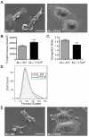
Fig. 1.
Fig. 1.
M−/−.Y721F macrophages display altered morphology. SEM images of M−/−.WT and M−/−.Y721F macrophages plated onto fibronectin-coated coverslips for 2 days and either kept in the continuous presence of CSF-1 (A) or incubated without growth factor overnight prior to stimulation with 120 ng/ml CSF-1 for 3 minutes (E) before fixation. Scale bars: 20 μM. M−/−.WT and M−/−.Y721F macrophages plated onto fibronectin-coated coverslips and grown in the continuous presence of CSF-1 were also fixed and stained for F-actin before cell outline tracing to measure the cell footprint area (B) and elongation ratio (maximum length:maximum width) (C). Data are means±s.e.m. (n≥40). ***P<0.001. (D) Forward scatter analysis of M−/−.WT and M−/−.Y721F cell size (FACSCantoII, BD Biosciences).
Fig. 2.
Fig. 2.
Paxillin and Pyk2 phosphorylation, and the incorporation of paxillin into adhesion structures, are reduced in M−/−.Y721F macrophages. (A) M−/−.WT and M−/−.Y721F macrophages were plated onto fibronectin-coated coverslips and incubated without growth factor overnight before stimulation with 120 ng/ml CSF-1 for 15 minutes followed by fixation, staining and examination by TIRF microscopy for adhesion structures rich in paxillin Y118-P. Scale bar: 20 μM. Lysates from CSF-1-starved M−/−.WT and M−/−.Y721F macrophages stimulated with 120 ng/ml CSF-1 for the indicated times were subjected to paxillin (B) or Pyk2 (C) immunoprecipitation (IP) then immunoblotted (WB) for the indicated proteins.
Fig. 3.
Fig. 3.
Loss of CSF-1R Y721-P signaling increases expression of the adhesion-regulating PTPϕ. (A) SEM images of M−/−.WT macrophages grown in the continuous presence of either GM-CSF or CSF-1 for 1 week before plating on fibronectin-coated coverslips for 2 days. Scale bar: 20 μM. (B) M−/−.WT and M−/−.Y721F cell lysates were examined by SDS-PAGE and immunoblotting for the indicated proteins. (C) Levels of mRNA encoding FAK, Pyk2 and PTPϕ were examined in M−/−.WT and M−/−.Y721F macrophages cultured in the continuous presence of CSF-1 by qRT-PCR analysis. Results are expressed as fold-change in M−/−Y721F cell gene expression compared with that in M−/−.WT cells. The data are means±s.e.m. for three independent experiments. *P<0.05.
Fig. 4.
Fig. 4.
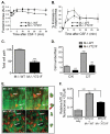
CSF-1R Y721-P signaling regulates macrophage spreading, motility and promotion of carcinoma cell invasion. CSF-1-stimulated M−/−.WT (▴) and M−/−.Y721F (▪) macrophages plated onto glass-bottomed dishes were examined by time-lapse video microscopy and their cell outlines traced. The mean (±s.e.m.) total footprint area (A) and relative (%) increase in footprint area (B) for 30 minutes after CSF-1 (120 ng/ml) addition is shown (n≥40). (C) Total path length was measured by centroid analysis at 2.5-minute intervals up to 1 hour after CSF-1 stimulation and means±s.e.m. calculated for 10 cells each. (D) Boyden chamber analysis of CSF-1-stimulated chemokinetic (C/K) and chemotactic (C/T) motility is shown. The data are normalized to the M−/−.WT C/K result and are means±s.e.m. for a representative experiment (n=3). (E) M−/−.WT and M−/−.Y721F cells labeled with Cell Tracker Red (red) were injected into GFP-expressing MTLn3 xenograft carcinomas (green) 24 hours prior to imaging by multiphoton microscopy for 45 minutes. Scale bar: 20 μM. Two M−/−.WT and M−/−.Y721F cell (arrows) outlines in the first (green) and last (red) frame were overlaid to evaluate cell motility in vivo. (F) CFP-expressing MTLn3 carcinoma cells were either cultured alone or co-cultured with M−/−.WT or M−/−.Y721F cells and their capacity to invade more than 20 μM into overlaying collagen after 24 hours measured. The data are means±s.e.m. **P<0.01; ***P<0.001.
Fig. 5.
Fig. 5.
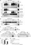
Mutation of CSF-1R Y721 abrogates its association with p85 PI3K, but not PLCγ, and induces PI3K expression. CSF-1-starved M−/−.WT and M−/−.Y721F cells were stimulated with CSF-1 for the indicated times and lysates subjected to CSF-1R (A), p85 PI3K (B), PY100 (C), PLCγ2 (D) and PLCγ1 (E) immunoprecipitation (IP). Eluted proteins were subjected to SDS-PAGE and immunoblotting (WB) for the indicated proteins. WCL, whole-cell lysates. (F) qRT-PCR analysis of mRNA encoding p85 PI3K and PLCγ2 in M−/−.WT and M−/−.Y721F macrophages grown continuously in CSF-1. Results are the fold-change in M−/−Y721F cell gene expression compared with that in M−/−.WT cells (means±s.e.m. for three independent experiments). *P<0.05. (G) CSF-1-starved M−/−.WT cells were treated with either 100 nM wortmannin or DMSO for 30 minutes before CSF-1 stimulation for the indicated times. F-actin-rich ruffles were visualized with Alexa-Fluor-568–phalloidin and quantified as described in the Materials and Methods.
Fig. 6.
Fig. 6.
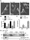
The CSF-1R Y721-P motif is necessary and sufficient for CSF-1-induced ruffling and chemotaxis, as well as association of p85 PI3K and CSF-1R. (A) SEM images of M−/−.WT, M−/−.Y721F and M−/−.Y3AB,Y721 macrophages plated onto fibronectin-coated coverslips. Cells were incubated without growth factor overnight before stimulation with 120 ng/ml CSF-1 for 3 minutes. Scale bar: 20 μM. Boyden chamber analysis of CSF-1-stimulated chemokinetic (C/K) and chemotactic (C/T) motility in M−/−.WT, M−/−.Y721F and M−/−.Y3AB,Y721 macrophages (B) or in M−/−.Y3AB and M−/−.Y3AB,Y721 macrophages (C). Data are means±s.e.m. for a representative experiment (n=3). **P<0.01; ***P<0.001. (D) CSF-1R immunoprecipitations (IP) of lysates from CSF-1-starved M−/−.WT, M−/−.Y721F, M−/−.Y544,Y559,Y807 and M−/−.Y3AB,Y721 cells, incubated without growth factor overnight before stimulation with 120 ng/ml CSF-1 for the indicated times, were examined for associated p85 PI3K following SDS-PAGE. The numbers below the blot indicate the relative levels of co-immunoprecipitated PI3K, as determined by densitometry.
Fig. 7.
Fig. 7.
PI3K association with the CSF-1R through Y721-P regulates Akt activation and PtdIns(3,4,5)P3 production. (A) M−/−.WT, M−/−.Y721F, M−/−.Y544,Y559,Y807 and M−/−.Y3AB,Y721 cells were stimulated with 120 ng/ml CSF-1 and their lysates examined for Akt S473 or T308 phosphorylation. (B) M−/−.WT, M−/−.Y721F and M−/−.Y3AB,Y721 cells plated onto fibronectin-coated coverslips were incubated without growth factor overnight before stimulation with 120 ng/ml CSF-1 for 0 or 15 seconds and were then subjected to simultaneous fixation and extraction with 3.7% paraformaldehyde, 0.1% glutaraldehyde and 0.15 mg/ml saponin to examine CSF-1-induced PtdIns(3,4,5)P3 production. Representative merged images consist of PtdIns(3,4,5)P3 (red) and phase-contrast (grayscale) images of M−/−.WT (i,iv), M−/−.Y721F (ii,v) and M−/−.Y3AB,Y721 (iii,vi) cells treated with CSF-1 for 0 (i–iii) or 15 (iv–vi) seconds. Arrows indicate PtdIns(3,4,5)P3-encircled vesicles and arrowheads point to CSF-1-induced PtdIns(3,4,5)P3 in lamellipodia. Images are representative of those in four independent experiments. Scale bar: 10 μM.
Fig. 8.
Fig. 8.
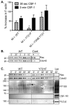
The Y721-P motif triggers signaling that regulates the first peak of actin polymerization and the association of an ~170 kDa phosphorylated protein with a Rac-GTP or Cdc42-GTP complex. (A) Relative (%) increase in cellular F-actin content in CSF-1-starved M−/−.WT, M−/−.Y721F and M−/−.Y3AB,Y721 cells stimulated with 120 ng/ml CSF-1 for either 30 seconds or 3 minutes. The data are means±s.e.m. (n=4). *P<0.05. (B) CSF-1-starved M−/−.WT cells were stimulated with 120 ng/ml CSF-1 for the indicated times and lysates subjected to GST–PAK–CRIB pull down for Rac-GTP and Cdc42-GTP followed by SDS-PAGE and immunoblotting for Rac and Cdc42. Positive and negative controls (Cont) were prepared and run at the same time. (C) CSF-1-starved M−/−.WT and M−/−.Y721F cells were stimulated with 120 ng/ml CSF-1 for 0 or 30 seconds and lysates (Lys) subjected to GST–PAK–CRIB pull down for Rac-GTP and Cdc42-GTP followed by SDS-PAGE and immunoblotting for the indicated proteins. The arrow indicates the ~170 kDa phosphorylated protein, seen in M−/−.WT samples only.

Similar articles

Cited by

References

    1. Abou-Kheir W., Isaac B., Yamaguchi H., Cox D. (2008). Membrane targeting of WAVE2 is not sufficient for WAVE2-dependent actin polymerization: a role for IRSp53 in mediating the interaction between Rac and WAVE2. J. Cell Sci. 121, 379-390 - PMC - PubMed
    1. Allen W. E., Jones G. E., Pollard J. W., Ridley A. J. (1997). Rho, Rac and Cdc42 regulate actin organization and cell adhesion in macrophages. J. Cell Sci. 110, 707-720 - PubMed
    1. Boocock C. A., Jones G. E., Stanley E. R., Pollard J. W. (1989). Colony-stimulating factor-1 induces rapid behavioural responses in the mouse macrophage cell line, BAC1. 2F5. J. Cell Sci. 93, 447-456 - PubMed
    1. Bourette R. P., Myles G. M., Choi J.-L., Rohrschneider L. R. (1997). Sequential activation of phosphatidylinositol 3-kinase and phospholipase C-γ2 by the M-CSF receptor is necessary for differentiation signaling. EMBO J. 16, 5880-5893 - PMC - PubMed
    1. Bunney T. D., Opaleye O., Roe S. M., Vatter P., Baxendale R. W., Walliser C., Everett K. L., Josephs M. B., Christow C., Rodrigues-Lima F., et al. (2009). Structural insights into formation of an active signaling complex between Rac and phospholipase C gamma 2. Mol. Cell 34, 223-233 - PubMed

Publication types

MeSH terms

Substances