

Optimization of electrical stimulation parameters for cardiac tissue engineering
- PMID: 21604379
- PMCID: PMC3117092
- DOI: 10.1002/term.377
Optimization of electrical stimulation parameters for cardiac tissue engineering
Abstract
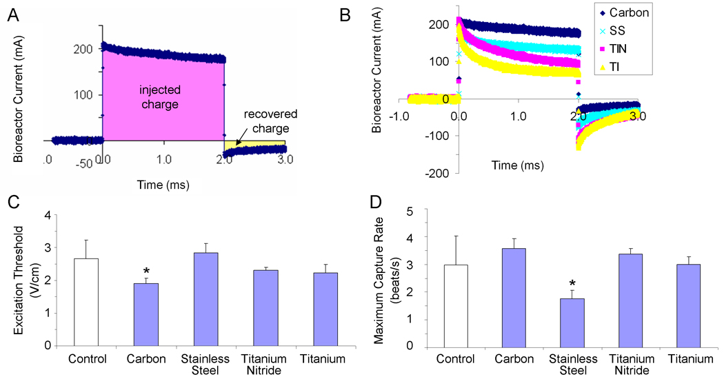
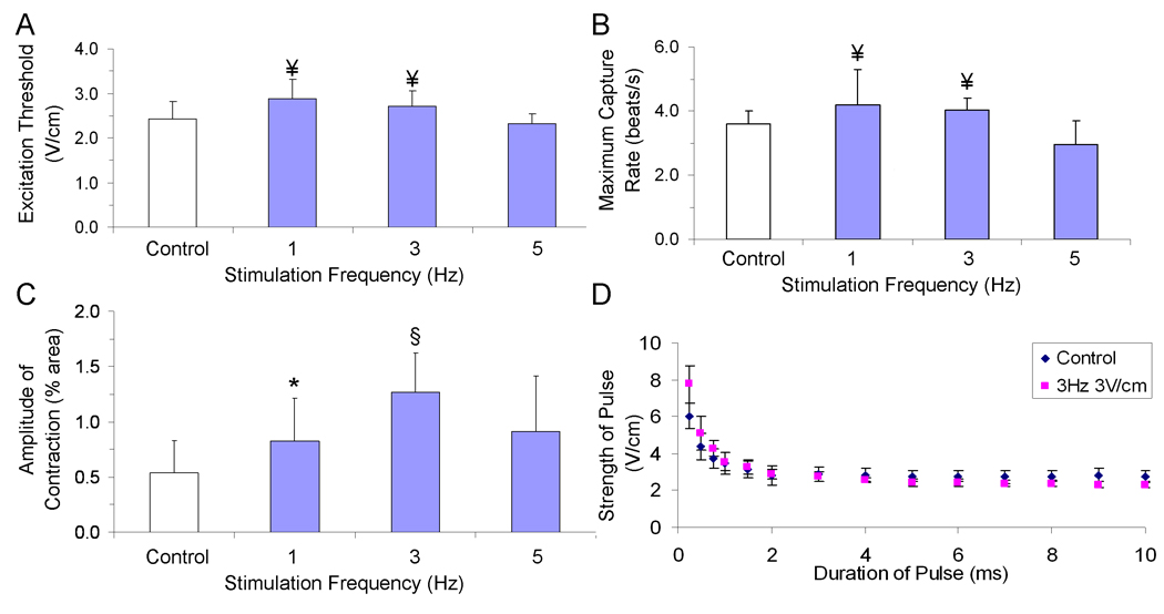
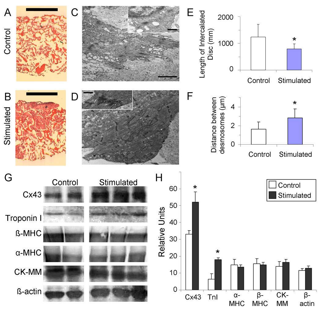
In vitro application of pulsatile electrical stimulation to neonatal rat cardiomyocytes cultured on polymer scaffolds has been shown to improve the functional assembly of cells into contractile engineered cardiac tissues. However, to date, the conditions of electrical stimulation have not been optimized. We have systematically varied the electrode material, amplitude and frequency of stimulation to determine the conditions that are optimal for cardiac tissue engineering. Carbon electrodes, exhibiting the highest charge-injection capacity and producing cardiac tissues with the best structural and contractile properties, were thus used in tissue engineering studies. Engineered cardiac tissues stimulated at 3 V/cm amplitude and 3 Hz frequency had the highest tissue density, the highest concentrations of cardiac troponin-I and connexin-43 and the best-developed contractile behaviour. These findings contribute to defining bioreactor design specifications and electrical stimulation regime for cardiac tissue engineering.
Copyright © 2011 John Wiley & Sons, Ltd.
Figures


Similar articles
-
Design of electrical stimulation bioreactors for cardiac tissue engineering.Annu Int Conf IEEE Eng Med Biol Soc. 2008;2008:3594-7. doi: 10.1109/IEMBS.2008.4649983. Annu Int Conf IEEE Eng Med Biol Soc. 2008. PMID: 19163486 Free PMC article.
-
Electric field stimulation integrated into perfusion bioreactor for cardiac tissue engineering.Tissue Eng Part C Methods. 2010 Dec;16(6):1417-26. doi: 10.1089/ten.TEC.2010.0068. Epub 2010 May 10. Tissue Eng Part C Methods. 2010. PMID: 20367291
-
Optical mapping of impulse propagation in engineered cardiac tissue.Tissue Eng Part A. 2009 Apr;15(4):851-60. doi: 10.1089/ten.tea.2008.0223. Tissue Eng Part A. 2009. PMID: 18847360 Free PMC article.
-
Electrical and mechanical stimulation of cardiac cells and tissue constructs.Adv Drug Deliv Rev. 2016 Jan 15;96:135-55. doi: 10.1016/j.addr.2015.07.009. Epub 2015 Jul 30. Adv Drug Deliv Rev. 2016. PMID: 26232525 Free PMC article. Review.
-
Electrical stimulation through conductive scaffolds for cardiomyocyte tissue engineering: Systematic review and narrative synthesis.Ann N Y Acad Sci. 2022 Sep;1515(1):105-119. doi: 10.1111/nyas.14812. Epub 2022 Jun 8. Ann N Y Acad Sci. 2022. PMID: 35676231 Free PMC article. Review.
Cited by
-
Versatile electrical stimulator for cardiac tissue engineering-Investigation of charge-balanced monophasic and biphasic electrical stimulations.Front Bioeng Biotechnol. 2023 Jan 4;10:1031183. doi: 10.3389/fbioe.2022.1031183. eCollection 2022. Front Bioeng Biotechnol. 2023. PMID: 36686253 Free PMC article.
-
Biomimetic cardiovascular platforms for in vitro disease modeling and therapeutic validation.Biomaterials. 2019 Apr;198:78-94. doi: 10.1016/j.biomaterials.2018.08.010. Epub 2018 Aug 4. Biomaterials. 2019. PMID: 30201502 Free PMC article. Review.
-
Electrical Stimulation and Cellular Behaviors in Electric Field in Biomedical Research.Materials (Basel). 2021 Dec 27;15(1):165. doi: 10.3390/ma15010165. Materials (Basel). 2021. PMID: 35009311 Free PMC article. Review.
-
Monophasic and biphasic electrical stimulation induces a precardiac differentiation in progenitor cells isolated from human heart.Stem Cells Dev. 2014 Apr 15;23(8):888-98. doi: 10.1089/scd.2013.0375. Epub 2014 Jan 24. Stem Cells Dev. 2014. PMID: 24328510 Free PMC article.
-
3D bioprinting in cardiac tissue engineering.Theranostics. 2021 Jul 6;11(16):7948-7969. doi: 10.7150/thno.61621. eCollection 2021. Theranostics. 2021. PMID: 34335973 Free PMC article. Review.
References
-
- Adams DS. A New Tool for Tissue Engineers: Ions As Regulators of Morphogenesis During Development and Regeneration. Tissue Eng Part A. 2008;14:1461–1468. - PubMed
-
- Bay BK. Texture correlation: A method for the measurement of detailed strain distributions within trabecular bone. J Orthop Res. 1995;13:258–267. - PubMed
-
- Berger HJ, Prasad SK, Davidoff AJ, et al. Continual electric field stimulation preserves contractile function of adult ventricular myocytes in primary culture. Am J Physiol Heart Circ Physiol. 1994;266:H341–H349. - PubMed
-
- Braeken D, Huys R, Jans D, et al. Local electrical stimulation of single adherent cells using three-dimensional electrode arrays with small interelectrode distances. Conf Proc IEEE Eng Med Biol Soc. 2009;2009:2756–2759. - PubMed
Publication types
MeSH terms
Grants and funding
LinkOut - more resources
Full Text Sources
Other Literature Sources
Research Materials
