. 2011 Jul 1;187(1):372-81.
doi: 10.4049/jimmunol.1100823. Epub 2011 May 20.

Human lung hydrolases delineate Mycobacterium tuberculosis-macrophage interactions and the capacity to control infection

Affiliations

Human lung hydrolases delineate Mycobacterium tuberculosis-macrophage interactions and the capacity to control infection

Jesús Arcos et al. J Immunol. .

Abstract

Pulmonary surfactant contains homeostatic and antimicrobial hydrolases. When Mycobacterium tuberculosis is initially deposited in the terminal bronchioles and alveoli, as well as following release from lysed macrophages, bacilli are in intimate contact with these lung surfactant hydrolases. We identified and measured several hydrolases in human alveolar lining fluid and lung tissue that, at their physiological concentrations, dramatically modified the M. tuberculosis cell envelope. Independent of their action time (15 min to 12 h), the effects of the hydrolases on the M. tuberculosis cell envelope resulted in a significant decrease (60-80%) in M. tuberculosis association with, and intracellular growth of the bacteria within, human macrophages. The cell envelope-modifying effects of the hydrolases also led to altered M. tuberculosis intracellular trafficking and induced a protective proinflammatory response to infection. These findings add a new concept to our understanding of M. tuberculosis-macrophage interactions (i.e., the impact of lung surfactant hydrolases on M. tuberculosis infection).


Figures

Figure 1
Figure 1
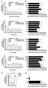
Basal expression of several hydrolase activities in ALF and human lung tissue. Hydrolase activities present in ALF (n = 8 in triplicate, mean ± SEM) (A) and human lung tissue (a representative experiment [mean ± SD]) of n = 3 in triplicate (B) were monitored using a colorimetric method based on the release of p-nitrophenol upon specific substrate cleavage. Results show the presence of several hydrolases with different levels of activity that have the potential to remodel the cell envelope of M. tuberculosis within the alveolar environment. 1, Acid phosphatase; 2, α-mannosidase; 3, α-galactosidase; 4, β-galactosidase; 5, α-glucosidase; 6, β-glucosidase; 7, α-xylosidase; 8, α-fucosidase; 9, arylsulfatase; 10, fatty acid esterase-I; 11, nonspecific esterase; 12, AlkP; 13. alkaline phosphodiesterase; 14, phospholipase C; 15, peroxidase; 16, α-rhamnosidase; 17, fatty acid esterase-II. See also Supplemental Table I.
Figure 2
Figure 2
Alveolar hydrolases modify the M. tuberculosis cell envelope. A, TLC analysis of hydrophobic fragments released from live M. tuberculosis upon surfactant hydrolase activity (TLC, chloroform/methanol, 96:4 v/v) is shown (n = 3). Treatments were: 1. 0.9% NaCl; 2. human monocyte lysate; 3. human AM lysate; 4. human ALF; and 5. 0.9% NaCl. B, EM of treated-M. tuberculosis. Morphologies of the bacterial surface were compared among groups in which the bacteria were treated with 0.9% NaCl (control), AlkP, AcP, Est, Mix (AlkP+AcP+Est), or ALF from two independent human donors (ALF#1 and ALF#3) before fixation. Lower-power electron micrographs were obtained at original magnification ×98,000. Scale bars, 100 nm. C, Car-bohydrate composition analysis of hydrolase-treated M. tuberculosis. M. tuberculosis (2 × 10⁷) was treated with 0.9% NaCl (control) or physiological concentrations of purified hydrolases (AlkP, AcP, Est, or Mix) or human ALF from a healthy donor. Alditol acetates obtained from treated-whole bacteria were further analyzed by GC/MS based on bacterial numbers. Shown are cumulative data of n = 3 each performed in duplicate (mean ± SEM). D, Carbohydrate composition analysis of soluble M. tuberculosis cell envelope components. M. tuberculosis-treated lysate fractions were converted to alditol acetates and analyzed by GC/MS based on equal amounts of protein (10 mg/ml). Shown are cumulative data of n = 4, each performed in duplicate (mean ± SEM): Ara, Man, myo-Inos, and Glc. Student t test, *p < 0.05, **p 0.005, ***p < 0.001.
Figure 3
Figure 3
Presence of ManLAM and TDM on the bacterial surface of hydrolase- and/or ALF-treated M. tuberculosis. Whole bacterial ELISA using live treated M. tuberculosis and anti-LAM mAb CS-35 (for Man-LAM) (A, B) or anti-TDM polyclonal Ab (for TDM) (C, D). A and B are representative experiments of n = 3, each performed in triplicate (mean ± SD); Student t test, treatment versus control. C and D are overall data from n = 3, each performed in trip-licate (mean ± SEM). One-way ANOVA followed by post-Tukey's multiple comparison test, *p < 0.05; **p < 0.005; ***p < 0.001, treatment versus control. ManLAM (5 μg) or TDM (5 μg) were used aspositive control. Buffer: 0.9% NaCl. *p < 0.05, **p < 0.005, ***p < 0.001. Mix, AlkP+AcP+Est.
Figure 4
Figure 4
Association of treated-M. tuberculosis with human macrophages. Single-cell suspensions of M. tuberculosis (2 × 10⁷) were incubated with 0.9% NaCl (control), ALF in 0.9% NaCl, commercially available human AlkP (1.414 U/ml 0.9% NaCl) or AcP (0.5 U/ml 0.9% NaCl), or a hydrolase mixture (AlkP + AcP + Est [Est at 0.52 U/ml 0.9% NaCl]) for 15 min, 1, 3, 12, and 24 h (the latter not shown). Infections (using bacilli counted after treatment and washing) and cell-association studies using human macrophage mono-layers on coverslips were performed at an MOI of 10:1. A, A representative experiment for each length of hydrolase exposure performed in triplicate (mean ± SD). B, Overall data showing the percent association decrease for each length of hydrolase exposure studied (mean ± SEM); n = 2 for all the treatments except for 12 h treatment, for which n = 4 for AlkP, AcP, Mix, and ALF#1 treatments, n = 3 for Est and ALF#2 treatments, and n = 1 for ALF#3 treatment. ALF#1, ALF#2, and ALF#3 are ALFs obtained from three independent donors. C and D, Decrease in association with human macrophages is hydrolase treatment dependent. M. tuberculosis bacilli were treated with β-glucosidase, AlkP, or 0.9% NaCl. Data showing the number of M. tuberculosis associated with human macrophages (n = 3 in triplicate) (C) and the overall percent association decrease versus control (n = 3 in triplicate) (D) are depicted (mean ± SEM). Student t test, treatment versus control, *p < 0.05, **p < 0.005, ***p < 0.001.
Figure 5
Figure 5
Proinflammatory cytokine response induced by hydrolase- or ALF-treated M. tuberculosis. Human macrophage mono-layers were infected with 15 min- or 12 h-treated M. tuberculosis that was gently washed prior to addition to macrophages. TNF levels were measured from isolated supernatants by ELISA. TNF production induced by 15 min-treated M. tuberculosis (A, B) and TNF production induced by 12 h-treated M. tuberculosis (C, D) (mean ± SEM); n values are depicted in each graph. ALF#1, ALF#2, ALF#3, and ALF#4 are ALFs obtained from four independent donors. Student t test, treatment versus control, *p < 0.05, **p < 0.005, ***p < 0.001. nd, not detected.
Figure 6
Figure 6
Effects of human ALF and hydrolase activities on M. tuberculosis intracellular survival and trafficking in macrophages. M. tuberculosis bacilli were treated with 0.9% NaCl (control), ALF#1 (donor #1), a mixture of AlkP, AcP, and Est, or AlkP, AcP, and Est alone at relevant in vivo concentrations. Human macrophages were infected at an MOI of 1:1. M. tuberculosis survival was determined at the indicated intervals. A, A representative experiment in triplicate is shown (mean ± SD); Student t test, treatment versus control, *p < 0.05, **p < 0.005, ***p < 0.001. B, Cumulative data from three independent experiments each performed in triplicate (mean ± SEM); one-way ANOVA, Tukey-Post test, *p < 0.05, **p < 0.005, ***p < 0.001. Where no significance was reached by ANOVA analyses, direct pairwise comparisons between control and a specific hydrolase treatment were analyzed at each time point by Student t test, +p < 0.05. C, Fluorescence microscopy images of P–L fusion events in human macrophages in response to hydrolase- or ALF-treated M. tuberculosis. Human macrophages were adhered to glass coverslips and incubated with treated GFP-M. tuberculosis (MOI 10:1) for 2 h using a synchronized phagocytosis assay. Cell monolayers were fixed, permeabilized, and stained with anti-human CD63-PE–conjugated mouse anti-human IgG. Shown are merge images where CD63 positive compartments are red, GFP-treated-M. tuberculosis in unfused phagosomes are green (blue arrows), and those colocalized with CD63 are yellow (white arrows). P–L fusion was examined and enumerated via confocal microscopy, counting >350 P-L events per coverslip. Original magnification ×650. D, A representative experiment from n = 3 performed in triplicate (mean ± SD); Student t test treatment versus control, *p < 0.05. E, Overall percent P–L fusion increase from n = 3 in triplicate (mean ± SEM); Student t test treatment versus control, **p < 0.005, ***p < 0.001. ALF#1, ALF donor 1; C, Control (0.9% NaCl).

Similar articles

Cited by

References

    1. World Health Organization. Multidrug and Extensively Drug-Resistant TB (M/XDR-TB): 2010 Global Report on Surveillance and Response. WHO Press; Geneva, Switzerland: 2010.
    1. Schlesinger LS. The role of mononuclear phagocytes in tuberculosis. In: Lipscomb MF, Russell SW, editors. Lung macrophages and dendritic cells in health and disease. Marcel Dekker, Inc.; New York: 1997. pp. 437–480.
    1. Torrelles JB, Schlesinger LS. Diversity in Mycobacterium tuberculosismannosylated cell wall determinants impacts adaptation to the host. Tuberculosis (Edinb) 2010;90:84–93. - PMC - PubMed
    1. King RJ. Pulmonary surfactant. J Appl Physiol. 1982;53:1–8. - PubMed
    1. vanGolde LM. Synthesis of surfactant lipids in the adult lung. Annu Rev Physiol. 1985;47:765–774. - PubMed

Publication types

MeSH terms