Cooperative DNA and histone binding by Uhrf2 links the two major repressive epigenetic pathways
- PMID: 21598301
- PMCID: PMC3569875
- DOI: 10.1002/jcb.23185
Cooperative DNA and histone binding by Uhrf2 links the two major repressive epigenetic pathways
Abstract
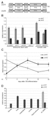
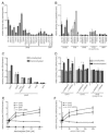
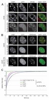
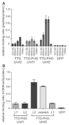
Gene expression is regulated by DNA as well as histone modifications but the crosstalk and mechanistic link between these epigenetic signals are still poorly understood. Here we investigate the multi-domain protein Uhrf2 that is similar to Uhrf1, an essential cofactor of maintenance DNA methylation. Binding assays demonstrate a cooperative interplay of Uhrf2 domains that induces preference for hemimethylated DNA, the substrate of maintenance methylation, and enhances binding to H3K9me3 heterochromatin marks. FRAP analyses revealed that localization and binding dynamics of Uhrf2 in vivo require an intact tandem Tudor domain and depend on H3K9 trimethylation but not on DNA methylation. Besides the cooperative DNA and histone binding that is characteristic for Uhrf2, we also found an opposite expression pattern of uhrf1 and uhrf2 during differentiation. While uhrf1 is mainly expressed in pluripotent stem cells, uhrf2 is upregulated during differentiation and highly expressed in differentiated mouse tissues. Ectopic expression of Uhrf2 in uhrf1(-/-) embryonic stem cells did not restore DNA methylation at major satellites indicating functional differences. We propose that the cooperative interplay of Uhrf2 domains may contribute to a tighter epigenetic control of gene expression in differentiated cells.
Copyright © 2011 Wiley-Liss, Inc.
Figures


Similar articles
-
S phase-dependent interaction with DNMT1 dictates the role of UHRF1 but not UHRF2 in DNA methylation maintenance.Cell Res. 2011 Dec;21(12):1723-39. doi: 10.1038/cr.2011.176. Epub 2011 Nov 8. Cell Res. 2011. PMID: 22064703 Free PMC article.
-
Molecular investigation of the tandem Tudor domain and plant homeodomain histone binding domains of the epigenetic regulator UHRF2.Proteins. 2022 Mar;90(3):835-847. doi: 10.1002/prot.26278. Epub 2021 Nov 19. Proteins. 2022. PMID: 34766381
-
Comparative biochemical analysis of UHRF proteins reveals molecular mechanisms that uncouple UHRF2 from DNA methylation maintenance.Nucleic Acids Res. 2018 May 18;46(9):4405-4416. doi: 10.1093/nar/gky151. Nucleic Acids Res. 2018. PMID: 29506131 Free PMC article.
-
The UHRF protein family in epigenetics, development, and carcinogenesis.Proc Jpn Acad Ser B Phys Biol Sci. 2022;98(8):401-415. doi: 10.2183/pjab.98.021. Proc Jpn Acad Ser B Phys Biol Sci. 2022. PMID: 36216533 Free PMC article. Review.
-
DNA methylation pathways and their crosstalk with histone methylation.Nat Rev Mol Cell Biol. 2015 Sep;16(9):519-32. doi: 10.1038/nrm4043. Nat Rev Mol Cell Biol. 2015. PMID: 26296162 Free PMC article. Review.
Cited by
-
UHRF2 regulates local 5-methylcytosine and suppresses spontaneous seizures.Epigenetics. 2017 Jul 3;12(7):551-560. doi: 10.1080/15592294.2017.1314423. Epub 2017 Apr 12. Epigenetics. 2017. PMID: 28402695 Free PMC article.
-
The nuclear protein UHRF2 is a direct target of the transcription factor E2F1 in the induction of apoptosis.J Biol Chem. 2013 Aug 16;288(33):23833-43. doi: 10.1074/jbc.M112.447276. Epub 2013 Jul 5. J Biol Chem. 2013. PMID: 23833190 Free PMC article.
-
Novel UHRF1-MYC Axis in Acute Lymphoblastic Leukemia.Cancers (Basel). 2022 Aug 31;14(17):4262. doi: 10.3390/cancers14174262. Cancers (Basel). 2022. PMID: 36077796 Free PMC article.
-
E3 ligase UHRF2 stabilizes the acetyltransferase TIP60 and regulates H3K9ac and H3K14ac via RING finger domain.Protein Cell. 2017 Mar;8(3):202-218. doi: 10.1007/s13238-016-0324-z. Epub 2016 Oct 14. Protein Cell. 2017. PMID: 27743347 Free PMC article.
-
Conserved linker regions and their regulation determine multiple chromatin-binding modes of UHRF1.Nucleus. 2015;6(2):123-32. doi: 10.1080/19491034.2015.1026022. Nucleus. 2015. PMID: 25891992 Free PMC article. Review.
References
-
- Abramoff MD, Magelhaes PJ, Ram SJ. Image processing with ImageJ. Biophotonics Int. 2004;11:36–42.
-
- Achour M, Fuhrmann G, Alhosin M, Ronde P, Chataigneau T, Mousli M, Schini-Kerth VB, Bronner C. UHRF1 recruits the histone acetyltransferase Tip60 and controls its expression and activity. Biochem Biophys Res Commun. 2009;390:523–528. - PubMed
-
- Arita K, Ariyoshi M, Tochio H, Nakamura Y, Shirakawa M. Recognition of hemi-methylated DNA by the SRA protein UHRF1 by a base-flipping mechanism. Nature. 2008;455:818–821. - PubMed
-
- Avvakumov GV, Walker JR, Xue S, Li Y, Duan S, Bronner C, Arrowsmith CH, Dhe-Paganon S. Structural basis for recognition of hemi-methylated DNA by the SRA domain of human UHRF1. Nature. 2008;455:822–825. - PubMed
Publication types
MeSH terms
Substances
LinkOut - more resources
Full Text Sources
Molecular Biology Databases
Miscellaneous

