. 2011 Aug;85(15):7622-33.
doi: 10.1128/JVI.00304-11. Epub 2011 May 18.

The internal Kaposi's sarcoma-associated herpesvirus LANA regions exert a critical role on episome persistence

Affiliations

The internal Kaposi's sarcoma-associated herpesvirus LANA regions exert a critical role on episome persistence

Erika De León Vázquez et al. J Virol. 2011 Aug.

Abstract

Kaposi's sarcoma-associated herpesvirus (KSHV) latency-associated nuclear antigen (LANA) is a 1,162-amino-acid protein that acts on viral terminal repeat (TR) DNA to mediate KSHV episome persistence. The two essential components of episome persistence are DNA replication prior to cell division and episome segregation to daughter nuclei. These functions are located within N- and C-terminal regions of LANA. N- and C-terminal regions of LANA are sufficient for TR DNA replication. In addition, N- and C-terminal regions of LANA tether episomes to mitotic chromosomes to segregate episomes to progeny cell nuclei. To generate a tethering mechanism, N-terminal LANA binds histones H2A/H2B to attach to mitotic chromosomes, and C-terminal LANA binds TR DNA and also associates with mitotic chromosomes. Here, we test the importance of the internal LANA sequence for episome persistence. We generated LANA mutants that contain N- and C-terminal regions of LANA but have most of the internal sequence deleted. As expected, the LANA mutants bound mitotic chromosomes in a wild-type pattern and also bound TR DNA as assayed by electrophoretic mobility shift assays (EMSA). The mutants mediated TR DNA replication, although with reduced efficiency compared with LANA. Despite the ability to replicate DNA and exert the chromosome and DNA binding functions necessary for segregating episomes to daughter nuclei, the mutants were highly deficient for the ability to mediate both short- and long-term episome persistence. These data indicate that internal LANA sequence exerts a critical effect on its ability to maintain episomes, possibly through effects on TR DNA replication.


Figures

Fig. 1.
Fig. 1.
Schematic diagram of KSHV LANA and LANA deletion mutants used in this investigation. Indicated are the proline-rich region (P), the aspartate- and glutamate-rich region (DE), the glutamine- and glutamate-rich region (Q) and the putative leucine zipper (LZ). The DE, Q, and LZ regions all contain repeat elements. The shaded region represents the N-terminal nuclear localization (NLS) signal. C-terminal LANA can also localize to nuclei but an NLS has not been precisely mapped. Amino acids 5 to 13 mediate chromosome association through interaction with histones H2A/H2B. Amino acids 996 to 1139 contain the TR DNA binding, self-association, and chromosome association functions. Capabilities for TR DNA binding, chromosome association, DNA replication, and episome persistence for each of the constructs are summarized at the right. Fractions are the numbers of G418-resistant cell lines containing episomes over the total number of G418-resistant cell lines assayed by Gardella analysis, and percentages are shown in parentheses.
Fig. 2.
Fig. 2.
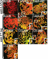
LANA and LANA deletion mutants associate with mitotic chromosomes. Control BJAB cells or BJAB cells stably expressing LANA, LANAΔ465–497, LANAΔ33–888, LANAΔ33–899, LANAΔ33–929, or LANAΔ33–949 were metaphase arrested with Colcemid and analyzed for LANA localization by confocal microscopy. LANA (green) was detected with anti-LANA antibody or anti-T7 epitope tag antibody. Chromosomes were counterstained with propidium iodide (red). The overlay of green and red generates yellow. Insets show enlargements of boxed chromosomes. Arrowheads indicate pericentromeric staining, and arrows indicate peritelomeric staining. Magnification, ×630.
Fig. 3.
Fig. 3.
Deletion of the internal LANA regions does not abolish DNA binding as detected by electrophoretic mobility shift assays (EMSA). Radiolabeled wild-type TR DNA (TR-13) probe (wt TR) containing the LANA high-affinity binding site was incubated with rabbit reticulocyte lysate (RRL) (lanes 1, 15, 29) or with in vitro-translated LANA (lanes 3, 17, 30), LANAΔ33–929 (lane 7), LANAΔ33–949 (lane 11), LANAΔ33–888 (lane 21), LANAΔ33–899 (lane 25), or LANAΔ465–497 (lane 31). Radiolabeled mutated TR DNA (Ti7) probe (mut. TR), which differs from TR-13 by a single base pair and abolishes the interaction with LANA, was incubated with RRL (lanes 2 and 16), LANA (lanes 4, 18), LANAΔ33–929 (lane 8), LANAΔ33–949 (lane 12), LANAΔ33–888 (lane 22), or LANAΔ33–899 (lane 26). Supershift analyses were performed after incubation with anti-T7 epitope tag antibody with LANA (lanes 5 and 19), LANAΔ33–929 (lane 9), LANAΔ33–949 (lane 13), LANAΔ33–888 (lane 23), or LANAΔ33–899 (lane 27). Incubation with IgG isotype-matched control for anti-T7 antibody did not result in supershifts for LANA (lanes 6 and 20), LANAΔ33–929 (lane 10), LANAΔ33–949 (lane 14), LANAΔ33–888 (lane 24), or LANAΔ33–899 (lane 28). Free probe is shown. Arrowheads indicate LANA-TR complexes or mutated LANA-TR complexes. Asterisks indicate supershifted complexes.
Fig. 4.
Fig. 4.
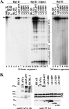
(A) Detection of p8TR replication mediated by LANA and LANA deletion mutants. Control BJAB cells, or BJAB cells stably expressing LANA or the LANA deletion mutants, were transfected with p8TR. Seventy-two hours posttransfection, low-molecular-weight DNA was isolated using the Hirt extraction method. Isolated DNA was digested with BglII (lanes 4 through 10) or BglII and DpnI (lanes 11 through 17), resolved in a 0.8% agarose gel, blotted onto nylon membrane, and detected by Southern blotting with radiolabeled TR probe. Lanes 1 through 3 contain the indicated amounts of p8TR plasmid digested with BglII. BJAB (lanes 4 and 11), LANA (lanes 5 and 12), LANAΔ465–497 (lanes 6 and 13), LANAΔ33–888 (lanes 7 and 14), LANAΔ33–899 (lanes 8 and 15), LANAΔ33–929 (lanes 9 and 16), and LANAΔ33–949 (lanes 10 and 17) cells are shown. The arrowhead indicates linearized p8TR. Nonreplicated (DpnI-sensitive) DNA and partially replicated DNA are indicated. Exposure to film at the left was for 12 h, and the panel at the right is the same blot for lanes 4 through 10, but after a 30-h exposure. This experiment is representative of two experiments. (B) Western blot for LANA and LANA mutants. BJAB cells, BCBL-1 PEL cells, or BJAB cells stably expressing LANA, LANAΔ465–497, LANAΔ33–888, LANAΔ33–899, LANAΔ33–929, or LANAΔ33–949 were assessed. LANA immune serum is more sensitive than T7 antibody, since it detects multiple LANA epitopes including within-repeat elements, so 150,000 cells per lane were used for this antibody, while 350,000 cells per lane were used for the T7 antibody blot. The vertical line indicates LANA, which includes full-length and also faster-migrating forms detected with LANA antibody. Asterisks indicate LANA and LANA mutants detected with T7 antibody. Nonspecific bands (NSB) are indicated.
Fig. 5.
Fig. 5.
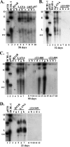
Internal LANA domains are critical for efficient long-term episome persistence. Control BJAB cells, or BJAB cells stably expressing LANA or the different LANA deletion mutants, were transfected with p8TR. Seventy-two hours posttransfection, transfected cells were seeded into microtiter plates at 1,000 cells/well and selected for G418 resistance. Gardella gel analysis detected p8TR episomes from G418-resistant cell lines expanded from the microtiter plates. Cells (∼1 × 10⁶ per lane) were loaded in Gardella gels. (A) Gardella gel containing naked p8TR plasmid (lane 1), BCBL-1 (KSHV-infected primary effusion cell line) (lane 2), p8TR-transfected G418-resistant BJAB cells (lanes 3 and 4), or p8TR-transfected G418-resistant BJAB cells expressing LANA (lanes 5 through 7) or LANAΔ465–497 (lanes 8 through 10). The Gardella gel analysis was done after 58 days of G418 selection. (B) Gardella gel containing BCBL-1 (lane 1), naked p8TR plasmid (lane 2), p8TR-transfected G418-resistant BJAB cells (lane 3), or p8TR-transfected G418-resistant BJAB cells expressing LANA (lanes 4 and 5), or LANAΔ33–929 (lanes 6 through 9). The Gardella analysis was done after 32 days of G418 selection. (C) Gardella gel containing BCBL-1 cells (lane 1), naked p8TR plasmid (lane 2), p8TR-transfected G418-resistant BJAB cells (lanes 3 and 4), or p8TR-transfected G418-resistant BJAB cells expressing LANA (lanes 5 through 7), LANAΔ33–888 (lanes 8 through 15), or LANAΔ33–899 (lanes 16 through 18). Vertical lines (lanes 10 and 18) indicate faint episomal signal. The Gardella gel analysis was done after 50 days of G418 selection. (D) Gardella gel containing naked p8TR plasmid (lane 1), BCBL-1 cells (lane 2), p8TR-transfected G418-resistant BJAB cells (lanes 3 and 4), and G418-resistant p8TR-transfected BJAB cells expressing LANA (lanes 5 through 7) or LANAΔ33–949 (lanes 8 through 12). The Gardella gel analysis was done after 22 days of G418 selection. Gel origin (O), BCBL-1 episomal (E) and linear (L) forms (due to KSHV lytic replication) and the p8TR circular, covalently closed (ccc) form are indicated.
Fig. 6.
Fig. 6.
Outgrowth of p8TR-transfected samples in bulk culture. BJAB (control) and BJAB cells stably expressing LANA and LANA deletion mutants were transfected with p8TR using Amaxa nucleofection. After transfection, cells were seeded in 6-well plates. Twenty-four hours posttransfection, cells were transferred to 175-cm2 flasks and seeded at 0.3 × 10⁶ cells/ml. Seventy-two hours posttransfection, cells were placed under G418 selection (600 μg/ml) at a concentration of 0.3 × 10⁶ cells/ml. Cells were then fed every other day and placed at 0.3 × 10⁶ cells/ml with each feeding. Cell concentration was recorded before the cells were cut back to 0.3 × 10⁶ cells/ml in fresh RPMI with G418. The data are an average of two experiments and include the experiment from Fig. 7.
Fig. 7.
Fig. 7.
Analysis of episome persistence at early time points indicates that the internal LANA domains exert a critical role. Control BJAB cells and BJAB cells stably expressing the different LANA constructs were transfected with p8TR and cultured as described in the legend to Fig. 6. Gardella cell analyses to detect the presence of episomal p8TR were performed at the indicated time points. Samples were resolved in 0.8% agarose gel followed by Southern blotting. DNA was detected using a 32P-labeled TR fragment probe, and the blot was exposed to film for the indicated times. In order to detect smaller amounts of DNA in control BJAB and in cells expressing LANA or LANA deletion mutants, twice as many cells were loaded per well (2 × 10⁶ cells) in Gardella gels at 12 days under selection and onwards. Naked plasmid p8TR DNA was loaded in indicated lanes as a control. (A) Episomal p8TR DNA at 24 h posttransfection. BCBL-1 (lane 1), p8TR plasmid (lane 2), BJAB cells (lane 3), BJAB cells expressing LANA (lane 4), LANAΔ465–497 (lane 5), LANAΔ33–888 (lane 6), LANAΔ33–899 (lane 7), LANAΔ33–929 (lane 8), or LANAΔ33–949 (lane 9). (B) Episomal p8TR DNA at 72 h posttransfection. BCBL-1 (lane 1), p8TR (lane2), BJAB (lane 4), BJAB cells expressing LANA (lane 5), LANAΔ465–497 (lane 6), LANAΔ33–888 (lane 7), LANAΔ33–899 (lane 8), LANAΔ33–929 (lane 9), or LANAΔ33–949 (lane 10). (C) Episomal p8TR DNA at 4 days under G418 selection (lanes 4 through 10) and 6 days under G418 selection (lanes 11 through 17). Pellets from cells at 4 days under G418 selection were frozen until the day of the Gardella analysis at 6 days under selection. BCBL-1 (lane 1), p8TR (lane 2), lane 3 is empty, control BJAB cells (lanes 4 and 11), and BJAB cells expressing LANA (lanes 5 and 12), LANAΔ465–497 (lanes 6 and 13), LANAΔ33–888 (lanes 7 and 14), LANAΔ33–899 (lanes 8 and 15), LANAΔ33–929 (lanes 9 and 16), and LANAΔ33–949 (lanes 10 and 17). (D) Episomal p8TR DNA at 8 days under G418 selection (lanes 4 through 10) and 10 days under G418 selection (lanes 11 through 17). Pellets from cells at 8 days under G418 selection were frozen until the day of the Gardella analysis at 10 days under selection. BCBL-1 (lane 1), p8TR (lane 2), lane 3 is empty, control BJAB cells (lanes 4 and 11), and BJAB cells expressing LANA (lanes 5 and 12), LANAΔ465–497 (lanes 6 and 13), LANAΔ33–888 (lanes 7 and 14), LANAΔ33–899 (lanes 8 and 15), LANAΔ33–929 (lanes 9 and 16), and LANAΔ33–949 (lanes 10 and 17). (E) Episomal p8TR DNA at 12 days under G418 selection (lanes 4 through 10) and 14 days under G418 selection (lanes 11 through 17). Pellets from cells at 12 days under G418 selection were frozen until the day of the Gardella analysis at 14 days under selection. BCBL-1 (lane 1), lane 2 is empty, p8TR (lane 3), control BJAB cells (lanes 4 and 11), and BJAB cells expressing LANA (lanes 5 and 12), LANAΔ465–497 (lanes 6 and 13), LANAΔ33–888 (lanes 7 and 14), LANAΔ33–899 (lanes 8 and 15), LANAΔ33–929 (lanes 9 and 16), and LANAΔ33–949 (lanes 10 and 17). The panel on the right is the same blot but is after a 3-day exposure to film to better show episomal p8TR in the LANA deletion mutants. (F) Episomal p8TR DNA at 16 days under G418 selection (lanes 4 through 10) and 18 days under G418 selection (lanes 11 through 17). Pellets from cells at 16 days under G418 selection were frozen until the day of the Gardella analysis at 18 days under selection. BCBL-1 (lane 1), p8TR (lane 2), lane 3 is empty, control BJAB cells (lanes 4 and 11), and BJAB cells expressing LANA (lanes 5 and 12), LANAΔ465–497 (lanes 6 and 13), LANAΔ33–888 (lanes 7 and 14), LANAΔ33–899 (lanes 8 and 15), LANAΔ33–929 (lanes 9 and 16), and LANAΔ33–949 (lanes 10 and 17). This figure is representative of two experiments.

Similar articles

Cited by

References

    1. An J., Sun Y., Rettig M. B. 2004. Transcriptional coactivation of c-Jun by the KSHV-encoded LANA. Blood 103:222–228 - PubMed
    1. Bajaj B. G., et al. 2006. KSHV encoded LANA upregulates Pim-1 and is a substrate for its kinase activity. Virology 351:18–28 - PubMed
    1. Ballestas M. E., Chatis P. A., Kaye K. M. 1999. Efficient persistence of extrachromosomal KSHV DNA mediated by latency-associated nuclear antigen. Science 284:641–644 - PubMed
    1. Ballestas M. E., Kaye K. M. 2001. Kaposi's sarcoma-associated herpesvirus latency-associated nuclear antigen 1 mediates episome persistence through cis-acting terminal repeat (TR) sequence and specifically binds TR DNA. J. Virol. 75:3250–3258 - PMC - PubMed
    1. Barbera A. J., Ballestas M. E., Kaye K. M. 2004. The Kaposi's sarcoma-associated herpesvirus latency-associated nuclear antigen 1 N terminus is essential for chromosome association, DNA replication, and episome persistence. J. Virol. 78:294–301 - PMC - PubMed

Publication types