. 2011 Jul;31(14):3019-28.
doi: 10.1128/MCB.05178-11. Epub 2011 May 16.

Chondrocyte-specific microRNA-140 regulates endochondral bone development and targets Dnpep to modulate bone morphogenetic protein signaling

Affiliations

Chondrocyte-specific microRNA-140 regulates endochondral bone development and targets Dnpep to modulate bone morphogenetic protein signaling

Yukio Nakamura et al. Mol Cell Biol. 2011 Jul.

Abstract

MicroRNAs (miRNAs) play critical roles in a variety of biological processes in diverse organisms, including mammals. In the mouse skeletal system, a global reduction of miRNAs in chondrocytes causes a lethal skeletal dysplasia. However, little is known about the physiological roles of individual miRNAs in chondrocytes. The miRNA-encoding gene, Mir140, is evolutionarily conserved among vertebrates and is abundantly and almost exclusively expressed in chondrocytes. In this paper, we show that loss of Mir140 in mice causes growth defects of endochondral bones, resulting in dwarfism and craniofacial deformities. Endochondral bone development is mildly advanced due to accelerated hypertrophic differentiation of chondrocytes in Mir140-null mice. Comparison of profiles of RNA associated with Argonaute 2 (Ago2) between wild-type and Mir140-null chondrocytes identified Dnpep as a Mir140 target. As expected, Dnpep expression was increased in Mir140-null chondrocytes. Dnpep overexpression showed a mild antagonistic effect on bone morphogenetic protein (BMP) signaling at a position downstream of Smad activation. Mir140-null chondrocytes showed lower-than-normal basal BMP signaling, which was reversed by Dnpep knockdown. These results demonstrate that Mir140 is essential for normal endochondral bone development and suggest that the reduced BMP signaling caused by Dnpep upregulation plays a causal role in the skeletal defects of Mir140-null mice.


Figures
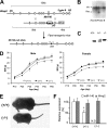
Fig. 1.
Fig. 1.
Generation of Mir140-null mice. (A) The Mir140 gene was replaced by an FRT-flanked, neomycin-resistant gene (Neo), which was subsequently removed by crossing mice with Flpe transgenic mice. (B) Southern blot analysis shows that an ES cell clone has both wild-type (Wt) and homologous recombinant (Rec) alleles. (C) PCR genotyping of mice using primers spanning FRT (see the small arrows in panel A). (D) Mir140-null mice show growth retardation in both male (left) and female (right) mice, whereas heterozygotes are indistinguishable from wild-type mice (n > 5). The body weight (BW) of Mir140-null mice was significantly lower than that of wild-type or heterozygous mice (n = 5; P < 0.05, t test) at all data points. (E) Picture of 3-week-old female mice. (F) miR-140 expression in primary rib chondrocytes is not detectable (N.D.) in Mir140-null mice, whereas miR-140 expression is reduced by approximately 50% in heterozygotes. The expression level of the host gene of Mir140, Wwp2, is not affected in Mir140-null mice.
Fig. 2.
Fig. 2.
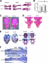
The longitudinal growth of endochondral bones is impaired in Mir140-null mice. Whole-mount Alizarin staining (A to D) shows the shortening of the indicated long bones (A), skull (C), and palate (D) of a 4-week-old male Mir140-null mouse. Brackets indicate palatal bones. (B) The lengths of the femur and tibia of 2-week-old Mir140-null mice are significantly smaller than those of wild-type littermates (n = 3; P < 0.05, t test). (E) Alizarin staining shows mild shortening of bones of the basal skull (right) of E17.5 Mir140-null embryos. BO, basioccipital bone; BS, basisphenoidal bone. (F) Hematoxylin and eosin-stained sections of the basisphenoidal bone (BS) of 5.5-day-old mice. The basisphenoidal bone (brackets) is shortened in Mir140-null mice. The bidirectional growth plates flanking this bone are slightly shortened. P, pituitary gland.
Fig. 3.
Fig. 3.
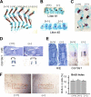
Mild acceleration of bone development in Mir140-null mice. Whole-mount skeletal staining (A to C) shows an advanced mineralization of the talus (arrow) in newborn Mir140-null hind limbs (A). The sizes of Alizarin red-stained mineralized areas of the talus (B) and forearms (C) are greater in Mir140-null mice than in wild-type or heterozygous littermates. Arrows indicate mineralization of the ulna. (D, E) The size of the hypertrophic region marked by Col10a1 (brackets) is greater in the talus at P0.5 (D) and the tibia at E14.5 (E) than in control animals, suggesting accelerated hypertrophic differentiation of chondrocytes. Brackets and black lines indicate the hypertrophic region. H/E, hematoxylin and eosin stain. (F) Growth plates of the Mir140-null tibia at P5.5 exhibit no significant proliferation defects in proliferating columnar chondrocytes.
Fig. 4.
Fig. 4.
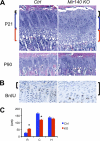
(A) Reciprocal change in the sizes of the resting and columnar zones of the Mir140-null growth plate. Hematoxylin and eosin-stained growth plates of the proximal tibia at P21 show an increase in the size of the resting zone (black brackets) and a modest decrease in the size of the columnar zone (blue brackets) in Mir140-null mice (KO) compared with those of control mice (Ctrl). This tendency is present but less appreciable in older mice (P90). (B) The resting zone (brackets), containing chondrocytes with low BrdU incorporation rates, is expanded in the Mir140-null growth plate. (C) The length of each layer was measured in 21-day-old control (wild-type and heterozygous mice) and Mir140-null littermates. The columnar zone (bars labeled C) was defined as the region containing columns formed by more than 5 chondrocytes per column. The resting (bars R) and hypertrophic (bars H) zones are defined as the cartilage area adjacent to the columnar zone (*, P < 0.05, n = 3, t test).
Fig. 5.
Fig. 5.
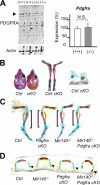
PDGFRA is not a physiologically important target of Mir140 in mouse chondrocytes. (A) PDGFRA expression is not upregulated in Mir140-null primary chondrocytes. Multiple bands detected by immunoblot analysis using anti-PDGFRA antibody in primary rib chondrocytes (left). The top two bands (arrowhead) migrate at the expected molecular masses (150 and 170 kDa). Pdgfra mRNA expression determined by qRT-PCR was not significantly altered in Mir140-null primary chondrocytes. N.S., not significant. (B) A chondrocyte-specific PDGFRA knockout (cKO) shows mild shortening of the longitudinal length of the skull and long bones. Skeletal preparation of the skull (left) and hind limbs (middle) at P10.5. Mineralization of the talus at P0.5 is slightly advanced in cKO mice (right). (C, D) Conditional ablation of Pdgfra does not rescue the skeletal defect of Mir140-null mice. (C) At P1.5, the shortening of the long bones of Mir140-null mice is not rescued by conditional Pdgfra ablation in chondrocytes. (D) The advanced mineralization of the hyoid horns (arrowheads) of 1.5-day-old Mir140-null mice was even augmented but not rescued in doubly mutant mice.
Fig. 6.
Fig. 6.
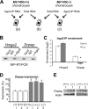
miR-140 targets Dnpep in mouse chondrocytes. (A) Experimental strategy. RNA was prepared from the Ago2-IP fraction and total RNA from wild-type and Mir140-null primary chondrocytes and subjected to expression profiling. RNA enriched in the Ago2-IP fraction was first identified in wild-type (a) and Mir140-null (c) chondrocytes; genes that were enriched in the Ago2-IP fraction in wild-type but not in Mir140-null chondrocytes were selected. Among them, those upregulated in Mir140-null chondrocytes (b) were subjected to further investigation. (B) RNA immunoprecipitation followed by RT-PCR (RIP-RT-PCR) shows the specific association between Hmga2 and Dnpep transcripts and Ago2 in wild-type cells. (C) Dnpep is associated with Argonaute 2 (Ago2) in wild-type but not in Mir140-null chondrocytes. Ago2 association was determined by comparing transcript levels of Ago2 immunoprecipitants with those of total RNA (Input in panel B). Transcript levels were expressed relative to that of GAPDH of each fraction. Ago2-IP enrichment ratios were calculated by dividing normalized transcript levels in the Ago2-IP RNA by those in total RNA. The relative enrichment of Dnpep in the Ago2-IP fraction is significantly less in Mir140-null chondrocytes, whereas Hmga2, a quintessential target of let-7 miRNAs, is enriched in the Ago2-IP fractions of both Mir140-null and wild-type chondrocytes (*, P < 0.05, n = 4, t test). (D, E) Dnpep is upregulated in Mir140-null primary rib chondrocytes both at the RNA level (D) and at the protein level (E).
Fig. 7.
Fig. 7.
Direct interaction between miR-140 and Dnpep suppresses Dnpep expression. (A) Coinjection of mouse or rat Dnpep rescues miR-140-induced palatal defects in zebrafish. Zebrafish embryos were injected with 3 nl of 25 μM miR-140 or 50 μg of miR-140 duplex solution with 1 ng of the indicated synthesized mRNA. The total numbers of analyzed fish are indicated in parentheses below the graph. The severity of the skeletal phenotypes was judged according to the criteria described previously (9). Fractions of fish with normal development were significantly greater in mouse or rat Dnpep-coinjected animals than in control animals (P < 0.05, χ2 test). (B) A major miR-140 binding site (BD) and its sequence predicted by STarMir are shown. The coding and untranslated regions are indicated by a gray box and lines, respectively. Underlines indicate seed sequence binding sites. (C) A luciferase reporter construct carrying the 45-nucleotide-long BD sequence, but not a mutated BD, shows a reduction in luciferase activity upon miR-140 cotransfection. The luciferase activity was normalized to that of cotransfected Renilla luciferase. (*, P < 0.05, n = 4, t test). (D) miR-140 transfection into ATDC5 cells reduces Dnpep expression.
Fig. 8.
Fig. 8.
Negative regulation of BMP activity by Dnpep. (A, B) Dorsalization in zebrafish overexpressing Dnpep. (A) Representative picture of dorsalized (white arrows) and normal (black arrow) embryos. Shortening of the tail and preservation of anterior structures of the head are observed at 30 h postfertilization. (B) About 4 to 6% of Dnpep-injected zebrafish show dorsalization (P < 0.05, χ2 test). The number of injected embryos per experimental group is indicated in parentheses above the graph. (C, D) Dnpep coinjection reduces the occurrence of ventralization caused by Bmp2b (P < 0.05, χ2 test). Numbers of injected animals are indicated in parentheses. Pictures were taken at 30 to 35 h postfertilization.
Fig. 9.
Fig. 9.
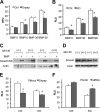
Dnpep dampens BMP signaling in mammalian cells. (A) BMP-responsive C2C12 cells were cotransfected with a BMP-responsive luciferase reporter (Id1-Luc), a Renilla luciferase (pRL-SV40) control, and an empty (control) or a Dnpep expression vector. Cells were treated with BMP-2 at the indicated concentrations (ng/ml) for 24 h. Relative luciferase activity normalized by that of Renilla luciferase was significantly reduced in Dnpep-overexpressing cells. (*, P < 0.05, n = 5, t test). RLU, relative light units. (B) Reduced BMP reporter activity in Mir140-null chondrocytes. Primary rib chondrocytes were transfected with Id1-Luc and treated with BMP-2 at the indicated concentrations for 24 h. Mir140-null chondrocytes show significantly lower normalized luciferase activities than those of the control (*, P < 0.05, n = 8, t test). (C, D) Smad phosphorylation (C) or the Smad4 level (D) is not affected in Mir140 deficiency. (E) Dnpep overexpression significantly reduced BMP reporter activity both in control and in Mir140-null (KO) chondrocytes (*, P < 0.05, n = 8, t test). Cells were cultured in the presence of 50 ng/ml of BMP-2 for 24 h. (F) An siRNA-mediated knockdown of Dnpep increased BMP reporter activity. siRNA duplexes were cotransfected to primary rib chondrocytes with Id1-Luc and pRL-SV40. Cells were cultured in the presence of 50 ng/ml of BMP-2 for 24 h. The Dnpep siRNA significantly increased the BMP reporter activity in Mir140-null chondrocytes (*, P < 0.05, n = 6, t test).

Similar articles

Cited by

References

    1. Ambros V., Chen X. 2007. The regulation of genes and genomes by small RNAs. Development 134:1635–1641 - PubMed
    1. Banegas I., et al. 2006. Brain aminopeptidases and hypertension. J. Renin Angiotensin Aldosterone Syst. 7:129–134 - PubMed
    1. Bartel D. P. 2009. MicroRNAs: target recognition and regulatory functions. Cell 136:215–233 - PMC - PubMed
    1. Beitzinger M., Peters L., Zhu J. Y., Kremmer E., Meister G. 2007. Identification of human microRNA targets from isolated argonaute protein complexes. RNA Biol. 4:76–84 - PubMed
    1. Blank U., et al. 2008. An in vivo reporter of BMP signaling in organogenesis reveals targets in the developing kidney. BMC Dev. Biol. 8:86. - PMC - PubMed

Publication types

MeSH terms