. 2011 May 16;193(4):633-42.
doi: 10.1083/jcb.201009069. Epub 2011 May 9.

NPHP4, a cilia-associated protein, negatively regulates the Hippo pathway

Affiliations

NPHP4, a cilia-associated protein, negatively regulates the Hippo pathway

Sandra Habbig et al. J Cell Biol. .

Abstract

The conserved Hippo signaling pathway regulates organ size in Drosophila melanogaster and mammals and has an essential role in tumor suppression and the control of cell proliferation. Recent studies identified activators of Hippo signaling, but antagonists of the pathway have remained largely elusive. In this paper, we show that NPHP4, a known cilia-associated protein that is mutated in the severe degenerative renal disease nephronophthisis, acts as a potent negative regulator of mammalian Hippo signaling. NPHP4 directly interacted with the kinase Lats1 and inhibited Lats1-mediated phosphorylation of the Yes-associated protein (YAP) and TAZ (transcriptional coactivator with PDZ-binding domain), leading to derepression of these protooncogenic transcriptional regulators. Moreover, NPHP4 induced release from 14-3-3 binding and nuclear translocation of YAP and TAZ, promoting TEA domain (TEAD)/TAZ/YAP-dependent transcriptional activity. Consistent with these data, knockdown of NPHP4 negatively affected cellular proliferation and TEAD/TAZ activity, essentially phenocopying loss of TAZ function. These data identify NPHP4 as a negative regulator of the Hippo pathway and suggest that NPHP4 regulates cell proliferation through its effects on Hippo signaling.


Figures
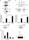
Figure 1.
Figure 1.
NPHP4 interacts with Lats1 and activates TAZ/YAP/TEAD transcription. (a) HEK293T cells were transiently transfected with the FLAG-tagged NPHPs NPHP1 (F.NPHP1) or NPHP4. After immunoprecipitation (IP) with FLAG (M2) beads, Western blot analysis revealed that endogenous Lats1 coprecipitated with NPHP4 but not with NPHP1. (b) Lysates from HEK293T cells transfected with either control vector (Input 1) or FLAG-tagged NPHP4 (Input 2 + 3) were subjected to immunoprecipitation using an anti-Lats1 (IP 1 and 2) or a control (anti-V5; IP 3) antibody. Western blot (WB) analysis showed that NPHP4 coprecipitated specifically with endogenous Lats1. (c) NPHP4 enhances the activity of the transcriptional coactivators TAZ and YAP. Plasmids encoding the indicated proteins or empty pcDNA6 vector were cotransfected in HEK293T cells together with the TEAD reporter plasmids as indicated in Materials and methods. Coexpression of NPHP4 increased the TAZ/YAP-dependent signaling to 156 and 138%, respectively (n = 3 for YAP and n = 5 for TAZ; *, P < 0.05). Expression of the indicated proteins was confirmed by Western blot analysis. (d) HEK293T cells were transfected with the indicated constructs. The chromatin immunoprecipitation assay revealed that NPHP4 enhances the binding of TAZ to the promoter region of CTGF, a known target gene of TAZ. Error bars represent SEM.
Figure 2.
Figure 2.
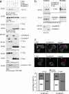
NPHP4 regulates the localization of TAZ. (a) HEK293T cells were transfected with the indicated constructs. Immunoprecipitation of TAZ or a control protein (Eps) with the FLAG antibody revealed that cotransfection of NPHP4, but not of NPHP1 or empty vector, decreased the interaction of TAZ with endogenous 14-3-3. Equal amounts of precipitated FLAG.TAZ were confirmed by staining the precipitates with anti-FLAG. Staining for TAZ in the cytoplasmic lysates used for immunoprecipitation (IP) showed reduced TAZ levels in the presence of NPHP4, whereas whole cell lysates (WCL), including the nuclear fraction, confirmed close to equal expression of TAZ. Staining for phosphorylation of the 14-3-3–binding motif of TAZ (phospho S89) revealed reduced phosphorylation of TAZ in the presence of NPHP4. (b) Cell fractionation experiments of HEK293T cells expressing the indicated proteins demonstrated that coexpression of NPHP4, but not NPHP1, increased the amount of TAZ in the nuclear compartment. Fibrillarin and 14-3-3 were used as nuclear and cytosolic marker proteins, respectively. (c) Breast epithelial cell MCF-10A was transfected with FLAG-tagged TAZ (F.TAZ) or FLAG-tagged YAP together with V5-tagged NPHP4 (V5.NPHP4) and stained with anti-FLAG and anti-V5 antibodies. In the absence of overexpressed NPHP4, F.TAZ/F.YAP showed a predominantly cytoplasmic localization (cells marked with arrowheads). In contrast, coexpression of V5.NPHP4 led to a highly significant increase in nuclear TAZ/YAP (bars, 10 µm; P < 0.01). WB, Western blot.
Figure 3.
Figure 3.
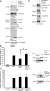
NPHP4 derepresses the inhibition of TAZ by Lats1. (a) NPHP4 abrogates the interaction of Lats1 and TAZ. FLAG-tagged Lats1 (F.Lats1) was coexpressed with GFP-tagged TAZ (GFP.TAZ) in the presence or absence of V5-tagged NPHP4 (V5.NPHP4) in HEK293T cells. After immunoprecipitation (IP) with FLAG (M2) beads, Western blot (WB) analysis revealed that the interaction of Lats1 and TAZ was abolished in the presence of NPHP4. FLAG.Eps served as a control protein. Both FLAG.Lats1 and FLAG.Eps were stained in the precipitates to control the comparable efficiency of the immunoprecipitation. (b) Overexpression of NPHP4 reduces the phosphorylation of TAZ by Lats1. HEK293T cells were transfected with the indicated constructs. Cell lysates were analyzed by Western blotting using the pYAP–Ser-127/pTAZ–Ser-89 antibody. (c) NPHP4 derepresses the inhibitory effect of Lats1 on TAZ signaling. Plasmids encoding the indicated proteins or empty pcDNA6 vector were cotransfected into HEK293T cells together with the TEAD reporter plasmids as indicated in Material and methods. As expected, Lats1 reduced TEAD/TAZ activity. Additional coexpression of NPHP4 reversed this effect (n = 4; *, P < 0.05). Expression of the indicated proteins was confirmed by Western blotting. (d) The Lats1-resistant mutant of TAZ (TAZ 4SA) could not be activated by NPHP4, suggesting that NPHP4 regulates TAZ signaling through interference with Lats1-dependent phosphorylation of TAZ (n = 3). Error bars represent SEM. WCL, whole cell lysate.
Figure 4.
Figure 4.
TAZ and NPHP4 influence cell proliferation and TAZ/TEAD target gene expression in MCF-7 cells. (a and b) MCF-7 cells were transfected with the indicated siRNAs. After 72 h, the cells were serum starved for 5 h followed by BrdU labeling for 30 min in the presence of serum. BrdU-positive cells were detected using an anti-BrdU antibody, and nuclei were counterstained using DAPI. The reduction of TAZ or NPHP4 leads to a decrease in cell proliferation, and the combined knockdown had no additional effect (n = 3; *, P < 0.05 as compared with the negative control; bars, 20 µM) (c) Knockdown of TAZ or NPHP4 led to a decreased expression of the TAZ/TEAD downstream target CTGF. 72 h after transfection of the indicated siRNAs into MCF-7 cells, the CTGF expression levels were analyzed using qPCR (n = 3; *, P < 0.05; **, P < 0.01). The knockdown of NPHP4 and TAZ was validated using qPCR (Fig. S3 a). Error bars represent SEM. hTAZ, human TAZ; hNPHP4, human NPHP4.
Figure 5.
Figure 5.
NPHP4 antagonizes the Hippo pathway. Active Hippo signaling leads to phosphorylation (P) and activation of the large tumor suppressor (LATS) kinase, which in turn phosphorylates YAP at Ser-127 and TAZ at Ser-89. This creates phosphoepitopes that are rapidly engaged by 14-3-3, leading to cytoplasmic retention of YAP and TAZ and the inhibition of their transcriptional activation potential. NPHP4 abrogates the interaction of large tumor suppressor with its substrates YAP and TAZ, which abolishes 14-3-3 binding and allows for nuclear translocation of YAP and TAZ.

Similar articles

Cited by

References

    1. Badouel C., Garg A., McNeill H. 2009. Herding Hippos: regulating growth in flies and man. Curr. Opin. Cell Biol. 21:837–843 10.1016/j.ceb.2009.09.010 - DOI - PubMed
    1. Berbari N.F., O’Connor A.K., Haycraft C.J., Yoder B.K. 2009. The primary cilium as a complex signaling center. Curr. Biol. 19:R526–R535 10.1016/j.cub.2009.05.025 - DOI - PMC - PubMed
    1. Bowers A.J., Boylan J.F. 2004. Nek8, a NIMA family kinase member, is overexpressed in primary human breast tumors. Gene. 328:135–142 10.1016/j.gene.2003.12.002 - DOI - PubMed
    1. Carter H., Samayoa J., Hruban R.H., Karchin R. 2010. Prioritization of driver mutations in pancreatic cancer using cancer-specific high-throughput annotation of somatic mutations (CHASM). Cancer Biol. Ther. 10:582–587 10.4161/cbt.10.6.12537 - DOI - PMC - PubMed
    1. Chan S.W., Lim C.J., Guo K., Ng C.P., Lee I., Hunziker W., Zeng Q., Hong W. 2008. A role for TAZ in migration, invasion, and tumorigenesis of breast cancer cells. Cancer Res. 68:2592–2598 10.1158/0008-5472.CAN-07-2696 - DOI - PubMed

Publication types

MeSH terms