. 2011 May 27;34(5):781-93.
doi: 10.1016/j.immuni.2011.03.020. Epub 2011 May 5.

Protective capacity of memory CD8+ T cells is dictated by antigen exposure history and nature of the infection

Affiliations

Protective capacity of memory CD8+ T cells is dictated by antigen exposure history and nature of the infection

Jeffrey C Nolz et al. Immunity. .

Abstract

Infection or vaccination confers heightened resistance to pathogen rechallenge because of quantitative and qualitative differences between naive and primary memory T cells. Herein, we show that secondary (boosted) memory CD8+ T cells were better than primary memory CD8+ T cells in controlling some, but not all acute infections with diverse pathogens. However, secondary memory CD8+ T cells were less efficient than an equal number of primary memory cells at preventing chronic LCMV infection and are more susceptible to functional exhaustion. Importantly, localization of memory CD8+ T cells within lymph nodes, which is reduced by antigen restimulation, was critical for both viral control in lymph nodes and for the sustained CD8+ T cell response required to prevent chronic LCMV infection. Thus, repeated antigen stimulation shapes memory CD8+ T cell populations to either enhance or decrease per cell protective immunity in a pathogen-specific manner, a concept of importance in vaccine design against specific diseases.


Figures

Figure 1
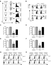
Figure 1. Secondary memory CD8+ T cells provide better protection against infections with LM, LCMV-Armstrong, and VacV, but decreased protection against MHV and LCMV clone 13
(A,B) Naïve B6 mice receiving either no cells (None) or Thy1.1 purified 2.5 × 10⁵ memory P14 TCR-tg CD8+ T cells (Primary or Secondary) were challenged with 1 × 10⁵ CFU of virulent LM-gp33. Bacterial burdens were analyzed in the liver on (A) day 3 or (B) day 5 post-infection. (C) Same as (A) except 5.0 × 10⁵ memory cells were transferred and mice were challenged with 2 × 10⁵ PFU of LCMV Armstrong. Viral titers were analyzed in the spleen on day 3 post-infection. (D) Same as (A) except 3.0 × 10⁵ memory cells were transferred and mice were infected intranasally with 1 × 10⁷ PFU of VacV-gp33. Viral titers were analyzed in the lung on day 3 post-infection. (E) Same as (C) except mice were infected intranasally with 1 × 10⁵ PFU of MHV-gp33 and titers were analyzed in the brain on day 7 post-infection. (F,G) Same as (A) except mice were challenged with 2 × 10⁶ PFU of LCMV clone 13. Viral titers were analyzed in the spleen on (F) day 3 and (G) day 10 post-infection. Dashed line indicates limit of detection (LOD). Statistical analyses employed the student’s t test. (see also Figure S1).
Figure 2
Figure 2. Secondary memory CD8+ T cells develop a unique phenotype of functional exhaustion during chronic LCMV infection
(A) Naïve mice receiving either no T cells (1° Response, Endogenous), 500 naïve P14’s (1° Response, P14 CD8+), 2.5 × 10⁵ primary (2° Response, P14 CD8+) or secondary memory P14’s (3° Response, P14 CD8+) were infected with 2 × 10⁶ PFU of LCMV clone 13. On day 7 post-infection, cells were obtained from blood of infected animals and stimulated ex vivo with gp33 peptide for 5 hours. IFNγ and TNFα positive cells were identified using intracellular staining. (B) Adoptive transfers and infections were performed as in (A). On day 25 post-infection, cells were obtained from spleen and P14 CD8+ T cells or endogenous gp33-specfic T cells were analyzed for expression of CD127 and the glycosylated isoform of CD43. (C–F) Adoptive transfers and infections were performed as in (A). On day 15 post-infection, expression of (C) PD-1, (D) LAG-3, (E) 2B4, and (F) CD160 was analyzed on the indicated CD8+ T cells in spleen. Representative histograms are shown in Figure S2. For (C–F), *P<0.001 compared to all other groups using the students t test. (G) Same as (A). Mice were then pulsed with BrdU on days 4–7 or 9–13. Representative BrdU incorporation profiles are shown from P14 CD8+ T cells from spleen. (H) Same as (G) except expression of Ki67 was assessed on day 7 or 13 post-infection. For (G) and (H), numbers indicate mean and std. dev. from 3 independent mice per group. (see also Figure S2).
Figure 3
Figure 3. CD62Lhi memory CD8+ T cells are equally protective against chronic viral infection regardless if they are primary or secondary
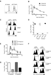
(A) 2 × 10⁶ total splenocytes from mice containing either primary (solid line histogram) or secondary (dashed line histogram) memory P14 CD8+ T cells were labeled with CFSE and were incubated with or without gp33 peptide for 60 hours. Representative CFSE profiles of Thy1.1 memory P14 CD8+ T cells with and without the addition of antigen are shown. (B) 2.5 × 10⁵ primary and secondary memory P14 CD8+ T cells (Thy1.1) were purified and adoptively transferred into naïve recipients (Thy1.2) which were subsequently infected with LCMV clone 13. Kinetics of the secondary or tertiary CD8+ T cell response were analyzed in the blood following infection using CD8+ and Thy1.1 staining to identify the adoptively transferred population. Error bars represent std. dev. of 3–5 mice at each time point. (C) Analysis of sorted CD62L high (CD62Lhi) and low (CD62Llo) expressing P14 CD8+ T cells from bulk primary and secondary memory populations. (D) 1.5 × 10⁵ cells from each of the groups in (C) were transferred into naïve recipients subsequently infected with LCMV clone 13. Viral titers were analyzed in the spleen on day 10 postinfection. (E) Same as (D) except kinetics of T cell expansion were monitored over the indicated time frame using CD8+ and Thy1.1 staining to identify the adoptively transferred populations. Error bars represent std. dev. of 3 samples for each time point. (F) Adoptive transfer of memory populations was performed as in (B) and recipient mice were subsequently infected with either LCMV clone 13 or LCMV Armstrong. On day 14 post-infection, TNFα production by IFNγ+ P14’s was assessed using intracellular stain following ex vivo stimulation with gp33 peptide. (G) Cumulative data of triplicate samples shown in (F). Data are representative of two independent experiments. (see also Figure S3).
Figure 4
Figure 4. Lymph node homing of memory CD8+ T cells is required for prevention of chronic LCMV infection
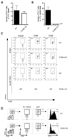
(A) 2.5 × 10⁵ primary memory P14 TCR-tg CD8+ T cells were transferred into either WT or Lta−/− mice, followed by infection with LCMV clone 13. Viral burden was analyzed from spleens on day 10 post-infection. Data are representative of two independent experiments. (B) 2.5 × 10⁵ primary or secondary memory P14 CD8+ T cells were transferred into Lta−/− mice and subsequently infected with virulent LM-gp33. Bacterial burden was analyzed in the liver on day 3 postinfection. (C) Equal numbers (2 × 10⁶) of WT (Thy1.1/1.1) and CD62L-deficient (CD62L-KO) (Thy1.1/1.2) primary memory P14 TCR-tg CD8+ T cells were transferred into naïve (Thy1.2/1.2) recipients. 48 hours later, organ-specific localization of both cell populations was determined. Representative histograms are shown in Figure S4H. (ILN, inguinal lymph node; CLN, cervical lymph node). (D,E) 2.5 × 10⁵ WT or CD62L–deficient primary memory P14 CD8+ T cells were purified and transferred into naïve recipients followed by infection with virulent LM-gp33. Bacterial burden was measured in the (E) spleen and (F) liver on day 3 post-infection. (F) Same as (D,E) except mice were infected with LCMV clone 13 and viral burden was analyzed in the spleen on day 10 post-infection. (see also Figure S4).
Figure 5
Figure 5. CD62L-deficient primary memory CD8+ T cells become activated in the spleen following LCMV clone 13 infection, but become functionally exhausted
(A,B) 2.5 × 10⁵ WT or CD62L-deficient (CD62L-KO) primary memory P14 TCR-tg CD8+ T cells were transferred into naïve B6 mice and subsequently infected with LCMV clone 13. Total numbers of P14 CD8+ T cells were determined in the (A) spleen or (B) inguinal lymph node on day 3 post-infection. (C) P14 CD8+ T cells from (A) were analyzed for expression of CD25 and CD69. (D) Adoptive transfers and infection was performed as in (A). On day 7 post-infection, cells were obtained from blood of infected animals and stimulated ex vivo with gp33 peptide for 5 hours. IFNγ and TNFα positive cells were identified using intracellular staining. Data are representative of two independent experiments.
Figure 6
Figure 6. Lymph node localization of memory CD8+ T cells results in enhanced viral clearance and sustained effector response during LCMV clone 13 Infection
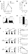
(A) 2.0 × 10⁵ WT or CD62L-deficient (CD62L-KO) primary memory CD8+ T cells were transferred into naïve recipients and subsequently infected with LCMV clone 13. On days 3, 5 and 7, total number of transferred cells was quantified in the inguinal lymph nodes. (B) Viral burden in the inguinal lymph node from (A) was determined on day 5 post-infection. (C,D) Same as (A) except on day 7 post-infection, lymph node cells were stimulated with gp33 peptide and TNFα production was analyzed on IFNγ+ cells using intracellular staining. (E) Equal numbers (1 × 10⁴) of WT (Thy1.1/1.2) or CD62L-deficient (Thy1.1/1.1) primary memory P14 TCR-tg CD8+ T cells were transferred into naïve B6 mice and subsequently infected with virulent LM-gp33, LCMV-Armstrong, or LCMV clone 13. On days 5, 6, 7, and 10, representation of each cell population was determined in the blood using Thy1.1 and Thy1.2 staining. Representative histograms from days 5 and 10 show representation of CD62L–deficient (Thy1.2-negative) and WT (Thy1.2-positive) P14 CD8+ T cells. Dashed line indicates input ratio. Error bars represent std. dev. of individual samples at each time point and data are representative of two independent experiments with 3 mice per group.
Figure 7
Figure 7. Blocking activated memory CD8+ T cell egress from lymph nodes following LCMV clone 13 infection results in impaired viral clearance
(A–C) 1.5 × 10⁵ primary memory P14 TCR-tg CD8+ T cells were transferred into naïve B6 mice and subsequently infected with LCMV clone 13. Mice were then treated with FTY720 or vehicle control on days 0, 2, and 4 post-infection. On day 6 post-infection, numbers of P14 CD8+ T cells were determined in the (A) blood, (B) spleen, or (C) inguinal lymph node. Representative dot plots for each organ are shown in Figure S7. For A–C, error bars represent std. dev. and statistical analysis was performed using the student’s t test. Data are representative of two independent experiments with 5 mice per group. (D) Same as (A) except mice were treated with FTY720 or vehicle control on days 0, 2, 4, 6, and 8 post-infection. On day 10 post-infection, viral burden was determined in the spleens of infected animals. Cumulative data from two independent experiments is shown. (see also Figure S5).

Similar articles

Cited by

References

    1. Arbones ML, Ord DC, Ley K, Ratech H, Maynard-Curry C, Otten G, Capon DJ, Tedder TF. Lymphocyte homing and leukocyte rolling and migration are impaired in L-selectin-deficient mice. Immunity. 1994;1:247–260. - PubMed
    1. Autran B, Carcelain G, Combadiere B, Debre P. Therapeutic vaccines for chronic infections. Science. 2004;305:205–208. - PubMed
    1. Badovinac VP, Messingham KA, Hamilton SE, Harty JT. Regulation of CD8+ T cells undergoing primary and secondary responses to infection in the same host. J Immunol. 2003;170:4933–4942. - PubMed
    1. Badovinac VP, Porter BB, Harty JT. Programmed contraction of CD8(+) T cells after infection. Nat Immunol. 2002;3:619–626. - PubMed
    1. Banks TA, Rouse BT, Kerley MK, Blair PJ, Godfrey VL, Kuklin NA, Bouley DM, Thomas J, Kanangat S, Mucenski ML. Lymphotoxin-alpha-deficient mice. Effects on secondary lymphoid organ development and humoral immune responsiveness. J Immunol. 1995;155:1685–1693. - PubMed

Publication types

MeSH terms