. 2011 Jun 15;186(12):7187-94.
doi: 10.4049/jimmunol.1100412. Epub 2011 May 4.

A functional role for Nlrp6 in intestinal inflammation and tumorigenesis

Affiliations

A functional role for Nlrp6 in intestinal inflammation and tumorigenesis

Grace Y Chen et al. J Immunol. .

Abstract

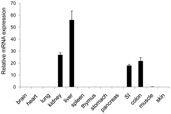
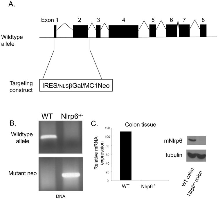
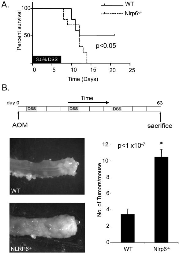
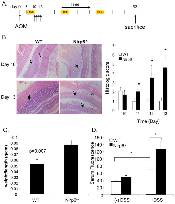
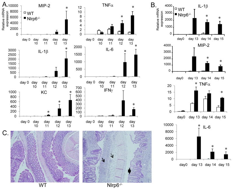
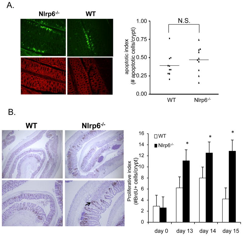
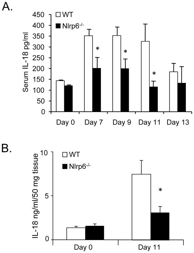
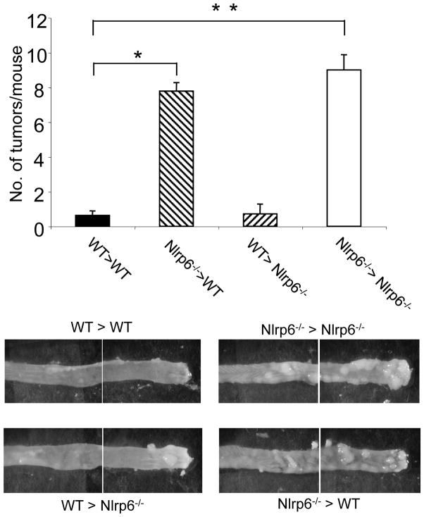
The nucleotide-binding oligomerization domain-like receptor (NLR) family member, Nlrp6, has been implicated in inflammasome signaling to activate caspase-1, which is essential for the production of mature IL-1β and IL-18. However, a function for Nlrp6 in vivo has never been demonstrated. Due to the relative high expression of Nlrp6 in intestinal tissue, we hypothesized that Nlrp6 has a role in intestinal homeostasis. Indeed, Nlrp6-deficient mice are more susceptible to chemically induced colitis as well as colitis-induced tumorigenesis than wild-type (WT) mice. Nlrp6-deficient mice exhibited significantly more inflammation within the colon than WT mice after dextran sulfate sodium treatment. Their inability to resolve inflammation and repair damaged epithelium as efficiently as WT mice resulted in prolonged increases in epithelial proliferative activity that likely underlie the increased propensity for tumors in these mice during chronic inflammation. We further show that the activity of Nlrp6 in hematopoietic cells is critical for protection against inflammation-related colon tumorigenesis. This study highlights the importance of NLR function in maintaining intestinal homeostasis to prevent the development of aberrant inflammation and tumor development within the colon.


Figures

Figure 1
Figure 1. Nlrp6 is relatively highly expressed in the intestine
mRNA expression of Nlrp6 relative to actin in various tissues isolated from at least 4 mice. Data represented by means +/−S.E.M.
Figure 2
Figure 2. Generation of Nlr6-deficient mice
A, Nlrp6−/− mice were generated by replacement of exon 1 and 2 of the Nlrp6 gene with the IRES/βGal/Neomycin resistance gene cassette. B, Genotype of Nlrp6−/− mice confirmed by PCR of genomic tail DNA using primers directed against Nlrp6, and the neomycin resistance gene. C, Confirmation of the absence of Nlrp6 expression within the colon of Nlrp6−/− mice on an mRNA (left) and protein (right) level. Nlrp6 mRNA was measured by quantitative real-time PCR and normalized with respect to actin. Nlrp6 protein expression in colon tissue extracts of wildtype (WT) and Nlrp6−/− mice was assessed by Western blot analysis with tubulin used as a loading control.
Figure 3
Figure 3. Nlrp6-deficient mice have increased susceptibility to chemically-induced colitis and inflammation-induced tumorigenesis
A, Ten age- and sex-matched wildtype (WT) and Nlrp6-deficient mice were treated with 7 days of 3.5% DSS and survival of mice was assessed. Data was analyzed by log-rank test. B, Age and sex-matched Nlrp6−/− (N=26 mice) and wildtype (N=25 mice) mice, were treated with AOM intraperitoneally followed by three cycles of 2% DSS. Three weeks after the last cycle of DSS, mice were sacrificed and tumors grossly counted with a stereomicroscope. Photographs are representative of tumors within the distal rectum of Nlrp6−/− and wildtype mice. Data is represented by means +/− S.E.M. *p <1 ×10⁻⁷ by Student’s t-test.
Figure 4
Figure 4. Increased inflammation and damage in Nlrp6-deficient mice
A, Representative photographs of jelly-rolled colons of Nlrp6−/− and wildtype (WT) mice sacrificed on day 10 and day 13 of the AOM/DSS tumor induction protocol under 250× magnification (left). Single arrows denote mucosal ulceration, dashed arrows point to submucosal edema, and thick arrow indicate regenerating crypts within the mucosa. Histologic scoring based on extent of mucosal ulceration, submucosal edema, and inflammatory cell infiltration was performed (N=5 mice/group/timepoint) treated with AOM/DSS and sacrificed on days 10 through 13 after the first round of DSS (right). C, Colons from sex- and age-matched Nlrp6−/− and wildtype (N=5 mice/group) were harvested on day 13, and weighed. Weights were normalized by length of colon. D, AOM/DSS-treated and untreated Nlrp6−/− and wildtype mice (N=5 mice/group) were given FITC-dextran by oral gavage on day 10. Serum was collected 4 hours later, and fluorescence measured by a fluorescent spectrophotometer. Data are expressed as means +/− S.E.M, *, p< 0.05 using Student’s t-test.
Figure 5
Figure 5. Increased proinflammatory mediator production and delayed resolution of inflammation in Nlrp6-deficient mice
A, mRNA was isolated from age- and sex-matched wildtype and Nlrp6-deficient on days 0 (N=3 mice/group), 10–13 (N=5 mice/group/timepoint) after treatment with AOM/DSS. mRNA expression of various proinflammatory mediators relative to β-actin was measured by quantitative real-time PCR. B, mRNA expression of proinflammatory mediators on days 13 to 15 after the first round of DSS in the AOM/DSS tumor induction protocol. Data expressed as means +/− S.E.M., *, p< 0.05 by Student’s t-test. C, Representative photographs of hematoxylin and eosin-stained colon sections from Nlrp6−/− and wildtype (WT) mice on day 15. Single arrow shows area of mucosal ulceration; dashed arrows point to submucosa edema. Thick arrows show regenerating crypts in a background of inflammation with inflammatory cell infiltration.
Figure 6
Figure 6. Increased epithelial proliferation in inflamed colons of Nlrp6-deficient mice
A, 10 age- and sex-matched mice were treated with AOM/DSS and sacrificed on day 8. Colons were harvested, jelly-rolled, formalin-fixed and paraffin-embedded. Levels of intestinal epithelial apoptosis was assessed by TUNEL assay using a fluorescent microscope. Representative photographs under 500× magnification of colon sections from Nlrp6−/− and wildtype mice (N=10 mice/group) stained for TUNEL activity (apoptotic cells labeled green, top) with corresponding sections counterstained with propidium iodide (red, bottom) (left). Apoptotic index represents number of apoptotic cells within the surface epithelium per crypt (~500 total crypts counted) (right). B, Age- and sex-matched Nlrp6−/− and wildtype mice (N=5 mice/group) were injected with BrdU intraperitoneally 2.5 hours prior to sacrifice on days 13–15 (N=5 mice/group/timepoint) and day 0 (N=3 mice/group). Proliferative activity of colonic epithelium was assessed by counting number of Brdu+ cells per crypt (~100 properly aligned crypts counted). Photographs are representative sections stained for BrdU at 250× magnification(top, left) and 500× (bottom, left). Arrow points to a regenerating crypt with disorganized architecture. Data expressed as means +/− S.E.M in bar graphs (right). *, p<0.05 by Student’s t-test. N.S., not significant.
Figure 7
Figure 7. Decreased Il-18 production in Nlrp6-deficient mice during inflammation-induced tumorigenesis
Serum was collected from age- and sex-matched untreated (N=3 mice/group) and AOM/DSS-treated Nlrp6−/− and wildtype mice on days 7 to 13 (N=5 to 9 mice/group/timepoint). IL-18 was measured by ELISA. B, Colon tissue protein extracts were made from untreated (N=4 mice/group) and AOM/DSS-treated mice (N=5 mice/group) sacrificed on day 11. IL-18 was measured by ELISA and normalized by colon weight. Data expressed as means +/− S.E.M., *, p<0.05 by Student’s t-test.
Figure 8
Figure 8. Nlrp6 function in hematopoietic cells is important for protection against inflammation-induced tumorigenesis
Bone marrow chimeras were generated by lethally-irradiating Nlrp6−/− and wildtype (WT) recipient mice and transplanting bone marrow by tail vein injection from either wildtype or Nlpr6−/− mice donor mice to generate 4 groups of mice: WT>WT (wildtype bone marrow into wildtype recipients) (N=8 mice), Nlrp6−/−>WT (N=9 mice), WT>Nlrp6−/− (N=7 mice), Nlrp6−/−>Nlrp6−/− (N=9 mice). Chimeric mice were treated with AOM/DSS and tumors counted after sacrifice (top). Data expressed as means +/−S.E.M., *,**p<0.05. Representative photographs of the mid-colon and distal colon sections for the four groups of chimeric mice after AOM/DSS treatment as viewed under a stereomicroscope (bottom).

Similar articles

Cited by

References

    1. Chen G, Shaw MH, Kim YG, Nunez G. NOD-like receptors: role in innate immunity and inflammatory disease. Annu Rev Pathol. 2009;4:365–398. - PubMed
    1. Watanabe T, Asano N, Murray PJ, Ozato K, Tailor P, Fuss IJ, Kitani A, Strober W. Muramyl dipeptide activation of nucleotide-binding oligomerization domain 2 protects mice from experimental colitis. J Clin Invest. 2008;118:545–559. - PMC - PubMed
    1. Bouskra D, Brezillon C, Berard M, Werts C, Varona R, Boneca IG, Eberl G. Lymphoid tissue genesis induced by commensals through NOD1 regulates intestinal homeostasis. Nature. 2008;456:507–510. - PubMed
    1. Zaki MH, Boyd KL, Vogel P, Kastan MB, Lamkanfi M, Kanneganti TD. The NLRP3 inflammasome protects against loss of epithelial integrity and mortality during experimental colitis. Immunity. 2010;32:379–391. - PMC - PubMed
    1. Chen GY, Shaw MH, Redondo G, Nunez G. The innate immune receptor Nod1 protects the intestine from inflammation-induced tumorigenesis. Cancer Res. 2008;68:10060–10067. - PMC - PubMed

Publication types

Substances