. 2011 Jul 1;286(26):23022-30.
doi: 10.1074/jbc.M111.225870. Epub 2011 May 2.

TIN2 protein dyskeratosis congenita missense mutants are defective in association with telomerase

Affiliations

TIN2 protein dyskeratosis congenita missense mutants are defective in association with telomerase

Dong Yang et al. J Biol Chem. .

Abstract

Dyskeratosis congenita (DC) is a progressive and heterogeneous congenital disorder that affects multiple systems and is characterized by bone marrow failure and a triad of abnormal skin pigmentation, nail dystrophy, and oral leukoplakia. One common feature for all DC patients is abnormally short telomeres and defects in telomere biology. Most of the known DC mutations have been found to affect core components of the telomerase holoenzyme. Recently, multiple mutations in the gene encoding the telomeric protein TIN2 have been identified in DC patients with intact telomerase genes, but the molecular mechanisms underlying TIN2 mutation-mediated DC remain unknown. Here, we demonstrate that ectopic expression of TIN2 with DC missense mutations in human cells led to accelerated telomere shortening, similar to the telomere phenotypes found in DC patients. However, this telomere shortening was not accompanied by changes in total telomerase activity, localization of TIN2, or telomere end protection status. Interestingly, we found TIN2 to participate in the TPP1-dependent recruitment of telomerase activity. Furthermore, DC mutations in TIN2 led to its decreased ability to associate with TERC and telomerase activity. Taken together, our data suggest that TIN2 mutations in DC may compromise the telomere recruitment of telomerase, leading to telomere shortening and the associated pathogenesis.


Figures

FIGURE 1.
FIGURE 1.
Ectopic expression of TIN2 DC mutants results in telomere shortening. A, schematic representation of TIN2 domain organization. The region frequently mutated in DC is shown. The amino acid positions studied here are underlined. B, Western blot (WB) analysis of HTC75 cells expressing FLAG-tagged wild-type or mutant TIN2. Control, empty vector. Anti-actin antibodies were used for loading controls. C, telomere restriction fragment analysis of cells from B. D, quantification of C to determine average telomere length. E, growth curve plot of cells from B.
FIGURE 2.
FIGURE 2.
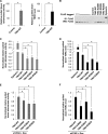
DC mutations in TIN2 do not affect the overall telomerase activity. A, TRAP assays were carried out with extracts from HTC75 cells expressing vector alone (Control) or FLAG-tagged wild-type and mutant TIN2. Heat-inactivated samples served as negative controls. B, the telomerase activities of cells from A were quantified. Error bars represent S.E. from three independent experiments. p values were calculated using Student's t test. C, the levels of TERC transcription in cells from A were determined by qPCR. The results were normalized to the transcriptional level of 18 S RNA. Error bars represent S.E. from triplicates of two independent experiments. p values were calculated using Student's t test.
FIGURE 3.
FIGURE 3.
DC mutations in TIN2 do not affect the expression of key telomeric proteins, their interaction with TIN2, or telomere end protection. A, the expression levels of the indicated endogenous telomeric proteins in control (empty vector) cells and cells expressing wild-type or mutant TIN2 were analyzed by Western blotting. Cells from early (PD18) and late (PD65) passages were examined. Arrowheads indicate FLAG-tagged wild-type and mutant TIN2. The asterisk indicates endogenous TIN2. short, short exposure; long, long exposure. B, TIN2 mutants maintained their ability to interact with TRF1, TRF2, and TPP1. FLAG-tagged wild-type TIN2 or mutants were transiently coexpressed with GST alone or with GST-tagged TRF1, TRF2, and TPP1 in 293T cells. GST fusion proteins were pulled down with glutathione beads, and the precipitated proteins were detected by Western blotting (WB). NR, nonrelevant sample. C, TIN2 mutants were targeted to the telomeres. Cells expressing GFP-tagged wild-type TIN2 or mutants were immunostained with anti-TRF2 antibodies (red). D, shown is the establishment of HT1080 cells with inducible TIN2 expression and stable TIN2 shRNA (shTIN2) knockdown. Expression of SFB-tagged wild-type or mutant TIN2 was induced with doxycycline (Dox). Control, empty vector. Anti-tubulin antibodies were used as a loading control. E, shown is the quantification of telomere dysfunction-induced focus assays performed with cells from D. Only cells with three or more 53BP1 foci that also co-localized with TRF2 foci were scored. Error bars represent S.E. (n = 3). p values were calculated using Student's t test. TIFs, telomere dysfunction-induced foci.
FIGURE 4.
FIGURE 4.
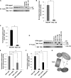
DC mutations in TIN2 compromise its ability to associate with the telomerase. A, wild-type TIN2 could bring down telomerase activity. FLAG-tagged wild-type TIN2 was immunoprecipitated from HTC75 cells with anti-FLAG antibodies, eluted with FLAG peptides, and used for Q-TRAP and TERC reverse transcription-PCR. B, anti-FLAG immunoprecipitates (IP) from HTC75 cells expressing wild-type or mutant TIN2 proteins were eluted with FLAG peptides for quantitative immunoblotting. TIN2 protein mixtures were serially diluted to obtain a calibration curve to normalize the protein amount in each immunoprecipitate. Dyskerin was included as a positive control for subsequent analysis. WB, Western blot. C, shown are the results from Q-TRAP analysis of the FLAG immunoprecipitates from B. The results were normalized based on the protein amount calculated in B. D, shown are the results from real-time qPCR analysis of the level of TERC in the FLAG immunoprecipitates from B. The results were normalized to the protein amount in the immunoprecipitates. E and F, examination of TIN2-associated telomerase activity and TERC levels in TIN2-inducible cells from supplemental Fig. S4. Cells stably expressing TIN2 shRNA (shTIN2) and doxycycline (Dox)-inducible SFB-tagged TIN2 proteins (supplemental Fig. S4) were treated with doxycycline, followed by immunoprecipitation with anti-FLAG antibodies. The immunoprecipitates were eluted with 3×FLAG peptides and used in Q-TRAP for telomerase activity (E) and qPCR for TERC levels (F) as described above. In A and C–F, error bars represent S.E. from triplicates of two independent experiments. p values were calculated using Student's t test. *, p < 0.05.
FIGURE 5.
FIGURE 5.
TIN2/TPP1 heterodimer recruits telomerase. A, anti-FLAG immunoprecipitates (IP) were prepared from HT1080 cells expressing inducible SFB-tagged wild-type TIN2 and TIN2-Δ90 and then eluted with 3×FLAG peptides for quantitative immunoblotting. The wild-type TIN2 immunoprecipitates were serially diluted to obtain a calibration curve to estimate the protein amount in each immunoprecipitate. WB, Western blot. B, Q-TRAP was carried out using the immunoprecipitates in A. The results were normalized to the protein amount. C, real-time qPCR was performed using the immunoprecipitates in A to determine TERC levels. The results were normalized to protein amount in the immunoprecipitates. D, anti-FLAG immunoprecipitates from HT1080 cells expressing inducible SFB-tagged wild-type or mutant TPP1 were eluted with 3×FLAG peptides for quantitative immunoblotting. The wild-type TPP1 immunoprecipitates were serially diluted to obtain a calibration curve to estimate the protein amount in each immunoprecipitate. E, Q-TRAP was carried out using the immunoprecipitates in D to determine telomerase activity. The results were normalized to the protein amount. F, real-time qPCR was performed using the immunoprecipitates in D to determine TERC levels. The results were normalized to 18 S RNA expression and protein amount in the immunoprecipitates. G, shown is a schematic model for TIN2-mediated telomerase recruitment and regulation. In B, C, E, and F, error bars represent S.E. from triplicates of three independent experiments. p values were calculated using Student's t test. *, p < 0.05.

Similar articles

Cited by

References

    1. Kirwan M., Dokal I. (2009) Biochim. Biophys. Acta 1792, 371–379 - PMC - PubMed
    1. Kirwan M., Dokal I. (2008) Clin. Genet. 73, 103–112 - PubMed
    1. Alter B. P., Baerlocher G. M., Savage S. A., Chanock S. J., Weksler B. B., Willner J. P., Peters J. A., Giri N., Lansdorp P. M. (2007) Blood 110, 1439–1447 - PMC - PubMed
    1. Vulliamy T. J., Knight S. W., Mason P. J., Dokal I. (2001) Blood Cells Mol. Dis. 27, 353–357 - PubMed
    1. Calado R. T., Young N. S. (2008) Blood 111, 4446–4455 - PMC - PubMed

Publication types

LinkOut - more resources