. 2011 May 27;34(5):729-40.
doi: 10.1016/j.immuni.2011.03.014. Epub 2011 Apr 28.

B7-h2 is a costimulatory ligand for CD28 in human

Affiliations

B7-h2 is a costimulatory ligand for CD28 in human

Sheng Yao et al. Immunity. .

Abstract

CD28 and CTLA-4 are cell surface cosignaling molecules essential for the control of T cell activation upon the engagement of their ligands B7-1 and B7-2 from antigen-presenting cells. By employing a receptor array assay, we have demonstrated that B7-H2, best known as the ligand of inducible costimulator, was a ligand for CD28 and CTLA-4 in human, whereas these interactions were not conserved in mouse. B7-H2 and B7-1 or B7-2 interacted with CD28 through distinctive domains. B7-H2-CD28 interaction was essential for the costimulation of human T cells' primary responses to allogeneic antigens and memory recall responses. Similar to B7-1 and B7-2, B7-H2 costimulation via CD28 induced survival factor Bcl-xL, downregulated cell cycle inhibitor p27(kip1), and triggered signaling cascade of ERK and AKT kinase-dependent pathways. Our findings warrant re-evaluation of CD28 and CTLA-4's functions previously attributed exclusively to B7-1 and B7-2 and have important implications in therapeutic interventions against human diseases.


Figures

Figure 1
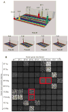
Figure 1. Identification of B7-H2-CD28 interaction by high throughput screening of a receptor array
(a) Screening of additional ligands for CD28 in a receptor array. CD28Ig was used as bait in the screening system. The 3-D illustration represents the result of a set of five 384-well plates. Each bar represents the total fluorescence intensity in the FL1 gate in each well of a 384-well plate. The Fc Receptors transfectants are used as positive controls. (b) Interactions among B7-CD28 family molecules. 293T cells were transfected with full length human B7-CD28 family genes as indicated on the x-axis. Fusion proteins were added to the culture as indicated on the y-axis to evaluate their bindings to the transfectants by CDS. Graphic view of individual wells is captured by 8200 CDS.
Figure 2
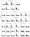
Figure 2. B7-H2 bound both CD28 and CTLA-4
(a–c) Specificity of B7-H2 bindings to CD28 and CTLA-4. 293T cells were transiently transfected with human full length B7-H2 (a), CD28 (b) or CTLA-4 (c) plasmids, and were subsequently stained by the indicated specific mAb or fusion proteins. In some experiments, additional mAb or fusion proteins were also included to examine the specificity of the binding. (d) Murine B7-H2 did not interact with murine CD28 and CTLA-4, while human B7-H2 interacts with murine ICOS, CD28 and CTLA-4. 293T cells were transiently transfected with mouse full length B7-H2, a hB7-H2IgV-mB7-H2 chimeric construct or human full length B7-H2 plasmids and were stained by the indicated fusion proteins. (e) Interactions of CD28 and CTLA-4 with their three ligands. To examine the competition between B7-H2 and B7-1 or B7-2 for binding CD28 and CTLA-4, B7-H2+ 293T cell transfectants were stained by CD28Ig and CTLA-4Ig, which were preincubated with excessive amount of B7-1Ig or B7-2Ig. Isotype-matched human Ig (Ctl Ig) was included as controls.
Figure 3
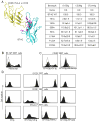
Figure 3. Interactions of B7-H2 with CD28, CTLA-4 and ICOS
(a) Left panel: Ribbon diagram of interacting model between B7-H2 and ICOS, CD28 or CTLA-4. The model was built by Pymol software (DeLano Scientific LLC.) based on crystal structure of B7-2-CTLA-4 (RCSB PDB 1i85). Right panel: binding site analysis of B7-H2 with its receptors by site-directed mutagenesis. Single point mutations were introduced into B7-H2 residues buried at the ligand-receptor interacting interface (shown in purple in ribbon diagram). B7-H2 plasmids (wild-type and mutants) were then transiently transfected into 293T cells and the expression of B7-H2 was confirmed by B7-H2 antibody staining. Binding of human ICOSIg, CD28Ig and CTLA-4Ig to B7-H2 mutants in transfected cells were measured by flow cytometry and normalized against 293T cells expressing wild-type B7-H2 (100%) and vector control (0%). The results were expressed as the percentage of binding to wild-type B7-H2. In addition to mean (n=3), 95% confidence intervals were given for each binding. (b–c) CTLA-4 competes with CD28 for B7-H2 binding. (b) CTLA-4Ig (Abatacept) was used to stain B7-H2+ 293T cells. (c) B7-H2Ig was pre-incubated with an excessive amount of Abatacept or control Ig for 15 min before staining CD28+ 293T cells. (d) Identification of mAbs that differentially abrogate B7-H2-CD28 and B7-H2-ICOS interactions. ICOS+, CD28+ or CTLA-4+ 293T cell transfectants were stained by B7-H2Ig or control (ctl) Ig. B7-H2 mAbs MIH12, 9F.8A4 or control mouse Ig, were included in the cultures to examine their blocking effects on B7-H2Ig binding.
Figure 4
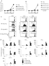
Figure 4. B7-H2 costimulation of human T cells through CD28
(a–b) B7-H2-CD28 interaction costimulates human T cell proliferation. Purified PBMC CD3+ T cells at 2.5x10⁵/wells were stimulated with immobilized human CD3 mAb (OKT3) and B7-H2Ig or control Ig (ctl Ig). (a) B7-H2 mAb (clone MIH12 or 9F.8A4) or control mouse Ig (ctl Ig) was added to block immobilized B7-H2Ig. (b) CD28 mAb (CD28.6) and/or ICOS mAb (C398.4A) or control Ig was added to the culture to block B7-H2 mediated costimulation. 3HTdR was added during the final six hours of culture. Similar results were also obtained in two additional experiments using different donors. (c) B7-H2-CD28 interaction promotes division of human T cells. Using the same condition as in (a), CFSE labeled CD3+ T cells were stimulated with immobilized anti-human CD3 and B7-H2Ig. B7-H2 mAb (clone MIH12 or 9F.8A4) was used to block coated B7-H2Ig. T cells were collected on day 4 and analyzed by a FACSCalibur flow cytometer. Average percentage of undivided T cell (n=3) and 95% confidence intervals are given. (d) B7-H2 costimulates cytokine production from human T cells through CD28. Purified CD4+ T cells at 2.5x10⁵/wells were stimulated by immobilized CD3 mAb and B7-H2Ig as described in (a). MIH12, 9F.8A4 or control mouse Ig (ctl Ig) was added to block B7-H2Ig. Supernatants were collected daily up to three days. Cytokine concentrations were measured by BD Cytometric Bead Array (CBA) human T helper-1 cell and T helper-2 cell (Th1-Th2 cell) cytokine kit and CBA IL-17A Flex set, and analyzed by FCAP Array software. Each data point is an average of three samples with standard deviation. Similar results were also obtained in two additional experiments using different donors.
Figure 5
Figure 5. B7-H2-CD28 interaction is important for T cell activation
(a–b) B7-H2-CD28 interaction enhances response of tetanus toxoid-specific memory T cells. (a) Monocyte-derived dendritic cells at 2.5x10⁴/well and purified autologous T cells at 3x10⁵/well were mixed together and incubated with tetanus toxoid at indicated concentration for five days. Blocking mAbs against CD28 (clone CD28.6), B7-H2 (clone MIH12 and 9F.8A4) or control mouse Ig at 5μg/ml was included in soluble form at the beginning of the culture. 3HTdR was added during the final sixteen hours of culture. * p<0.05. (b) Interferon gamma concentration was measured in day 5 supernatants by BD (CBA) Human Th1-Th2 cell cytokine kit and analyzed by FCAP Array software. Each data point is an average of samples with standard deviation. Similar results were also obtained in two additional experiments using different donors. (c–d) B7-H2-CD28 interaction promotes IFN-γ release in allogeneic T cell response. (c) Monocyte derived dendritic cells at 0, 1.25 x10⁴, 2.5x10⁴/well and purified allogeneic T cells at 3x10⁵/well were mixed together. Blocking mAbs against CD28 (clone CD28.6), B7-H2 (clone MIH12 and 9F.8A4) or control mouse Ig at 5μg/ml was included in soluble form at the beginning of the culture for five days. 3HTdR was added during the final sixteen hrs of culture. (d) Interferon gamma concentration was measured in day 5 supernatants by BD (CBA) Human Th1-Th2 cell cytokine kit and analyzed by FCAP Array software. Each data point is an average of three samples with standard deviation. Similar results were also obtained in two additional experiments using different donors.
Figure 6
Figure 6. B7-H2-CD28 interaction promotes Bcl-xL expression and downregulates p27kip1 in activated CD4+ T cells
Purified CD4+ T cells at 3.5x10⁵/well were stimulated with immobilized human CD3 mAb (0.1μg/ml) and 5μg/ml of immobilized (a) control Ig (ctl Ig), B7-1Ig, ICOS agonistic mAb (C398.4A), or (b and c) B7-H2Ig. In some wells, B7-H2 mAb (MIH12 or 9F.8A4), CD28 mAb (CD28.6), CTLA-4 mAb (14D3), ICOS mAb (C398.4A) or control mouse Ig was used to block B7-H2Ig mediated response. (b) Activated T cells were harvested on day 2 and day3, intracellularly stained by Bcl-xL mAb and analyzed by flow cytometry. (c) Activated T cells were harvested at 4 hours and 48 hours after activation. Cell lysates were prepared. 100μg total protein per sample was loaded and separated by 12% SDS-PAGE. The amount of phosphorylated GSK-3 (harvested at 4 hours) and p27kip1 (harvested at 48 hours) and beta-actin were determined by immunoblotting with respective antibodies. Beta actin was used as loading control.
Figure 7
Figure 7. B7-H2-CD28 interaction induces early activation of ERK and PI3K-AKT dependent pathways
(a) Purified native CD3+ T cells from wild-type or ICOS deficient mice at 3x10⁵/well were stimulated with immobilized 0.8μg/ml mouse CD3 mAb and 5μg/ml human or mouse B7-H2Ig or control Ig (ctl Ig). (b) In some wells, mouse T cells were first incubated with 5μg/ml mouse CD28 mAb (37.51) or control Ig for 15 minutes. After washing away unbound mAb, T cells were added to 96 well plates. 3HTdR was added during the final six hours of culture. Similar results were also obtained in two additional experiments. * p<0.05. (c) Purified mouse CD4+ T cells from wild-type, ICOS deficient or CD28 deficient mice were activated with 5μg/ml immobilized anti-CD3 for 3 days, and then rested for 3 days at the presence of 50IU/ml rmIL-2. T cells were subsequently harvested and added into 96 well plates pre-coated with 0.1μg/ml anti-CD3 plus 5μg/ml human B7-H2, mouse B7-1 or control Ig proteins. Cells were harvested at different time points as indicated. Cell lysates were prepared. 100μg total protein per sample was loaded and separated by 12% SDS-PAGE. The amount of phosphorylated ERK and AKT protein were determined by immunoblotting with phospho-specific antibodies (Thr202 and Tyr204 phospho-Erk1 and Erk 2 or Ser473 phospho-Akt). Membranes were subsequently striped and reprobed with antibodies against total ERK and AKT.

Similar articles

Cited by

References

    1. Appleman LJ, van Puijenbroek AA, Shu KM, Nadler LM, Boussiotis VA. CD28 costimulation mediates down-regulation of p27kip1 and cell cycle progression by activation of the PI3K/PKB signaling pathway in primary human T cells. J Immunol. 2002;168:2729–2736. - PubMed
    1. Boise LH, Minn AJ, Noel PJ, June CH, Accavitti MA, Lindsten T, Thompson CB. CD28 costimulation can promote T cell survival by enhancing the expression of Bcl-XL. Immunity. 1995;3:87–98. - PubMed
    1. Bossaller L, Burger J, Draeger R, Grimbacher B, Knoth R, Plebani A, Durandy A, Baumann U, Schlesier M, Welcher AA, et al. ICOS deficiency is associated with a severe reduction of CXCR5+CD4 germinal center Th cells. J Immunol. 2006;177:4927–4932. - PubMed
    1. Brodie D, Collins AV, Iaboni A, Fennelly JA, Sparks LM, Xu XN, van der Merwe PA, Davis SJ. LICOS, a primordial costimulatory ligand? Curr Biol. 2000;10:333–336. - PubMed
    1. Burmeister Y, Lischke T, Dahler AC, Mages HW, Lam KP, Coyle AJ, Kroczek RA, Hutloff A. ICOS controls the pool size of effector-memory and regulatory T cells. J Immunol. 2008;180:774–782. - PubMed

Publication types

MeSH terms