. 2011 Apr;9(4):e1001048.
doi: 10.1371/journal.pbio.1001048. Epub 2011 Apr 19.

Eps8 regulates hair bundle length and functional maturation of mammalian auditory hair cells

Affiliations

Eps8 regulates hair bundle length and functional maturation of mammalian auditory hair cells

Valeria Zampini et al. PLoS Biol. 2011 Apr.

Abstract

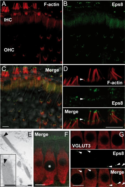
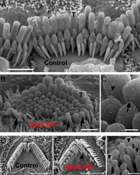
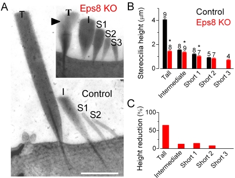
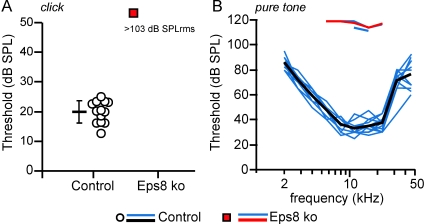
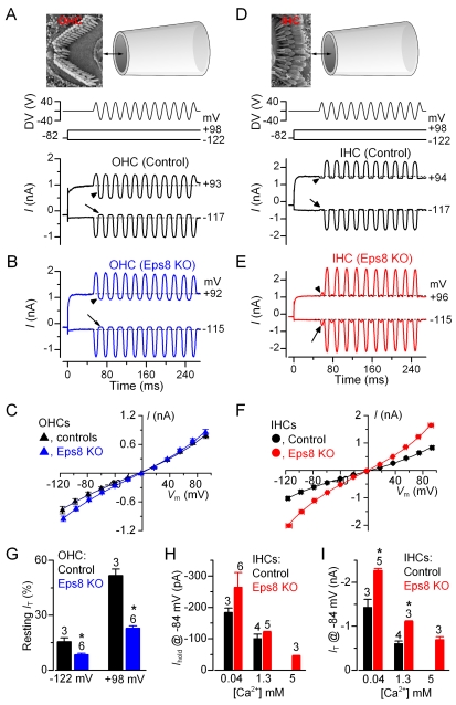
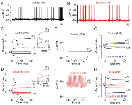
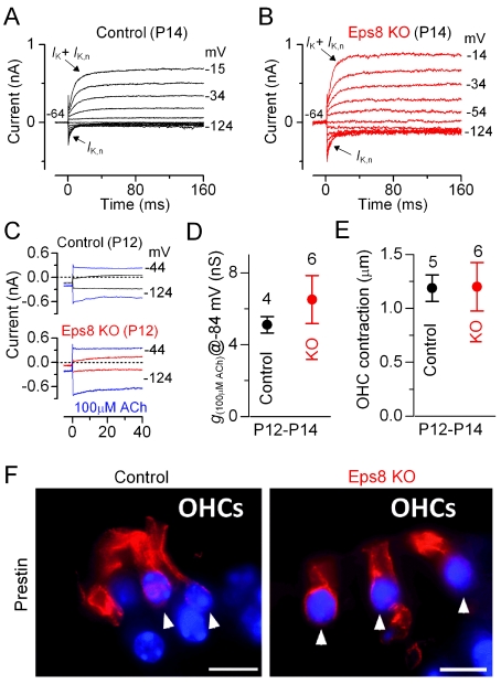
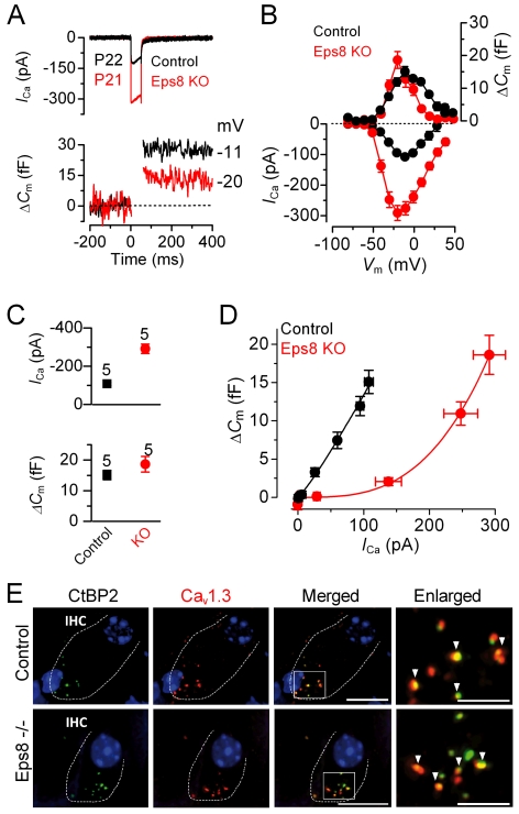
Hair cells of the mammalian cochlea are specialized for the dynamic coding of sound stimuli. The transduction of sound waves into electrical signals depends upon mechanosensitive hair bundles that project from the cell's apical surface. Each stereocilium within a hair bundle is composed of uniformly polarized and tightly packed actin filaments. Several stereociliary proteins have been shown to be associated with hair bundle development and function and are known to cause deafness in mice and humans when mutated. The growth of the stereociliar actin core is dynamically regulated at the actin filament barbed ends in the stereociliary tip. We show that Eps8, a protein with actin binding, bundling, and barbed-end capping activities in other systems, is a novel component of the hair bundle. Eps8 is localized predominantly at the tip of the stereocilia and is essential for their normal elongation and function. Moreover, we have found that Eps8 knockout mice are profoundly deaf and that IHCs, but not OHCs, fail to mature into fully functional sensory receptors. We propose that Eps8 directly regulates stereocilia growth in hair cells and also plays a crucial role in the physiological maturation of mammalian cochlear IHCs. Together, our results indicate that Eps8 is critical in coordinating the development and functionality of mammalian auditory hair cells.


Conflict of interest statement

The authors have declared that no competing interests exist.

Figures

Figure 1
Figure 1. Eps8 is localized at the hair cells stereociliary tips.
(A–C) F-actin containing stereocilia (A) and Eps8 (B) staining in IHCs and OHCs from P45 mice using confocal microscopy. Merged images are shown in (C) together with the DIC image. Note that Eps8 localizes at the tip of stereocilia (red: F-actin staining). (D) High resolution image of IHC stereocilia showing that Eps8 is present in the tips of both taller and shorter (arrowhead) stereocilia. Scale bars in (A–D) are 5 µm. (E) TEM immunogold labeling showing the stereociliary localization of Eps8 in a P35 IHC (inset shows this at higher magnification). Scale bars: 200 nm. (F) Immunostaining of IHCs from P16 mice using confocal microscopy. Image is a single section at the level of the IHC nuclei. Red indicates the IHC marker VGLUT3 and green Eps8. Asterisks denote IHC nuclei. Note that some stereociliary Eps8 staining is evident (top of the image) due to the angle of the tissue. (G) Magnified images of IHCs showing that punctate Eps8 labeling was also present in the cell cytoplasm (arrowheads). Scale bars in (F) and (G): 10 µm.
Figure 2
Figure 2. Hair bundle morphology in hair cells from adult Eps8 mice.
(A) Scanning electron micrograph (SEM) showing the typical hair bundle structure in control adult mouse P18 IHCs. Note that, generally, IHC hair bundles are composed of three rows of stereocilia. The tall stereocilia are indicated by T. Scale bar: 2 µm. (B) SEM showing that tall stereocilia (T) appear to be truncated in adult Eps8 knockout mice. Arrows indicate taller stereocilia oriented opposite to those positioned in the direction of bundle sensitivity. Moreover, additional rows of short stereocilia (three in this case) are present compared to wild type mice. Scale bar: 1 µm. (C) Higher magnification micrograph showing a tip link (arrow) and tip-link remnant (arrowhead) in an IHC from an Eps8 knockout mouse. Scale bar: 250 nm. (D–F) Outer hair cell hair bundle morphology in Eps8 mice. SEM showing the typical hair bundle structure of a second row (from the modiolus) OHC from the middle region of a normal wild type adult mouse cochlea (panel D). The precisely arranged rows of stereocilia with well defined height increments can be seen. (E) A comparable hair bundle from an Eps8 adult mutant mouse. Note that all the stereocilia in equivalent rows appear shorter, and the shortest row has missing stereocilia. Additional rows of stereocilia are also present (arrowhead). Scale bars: 2.5 µm. (F) Detail of the hair bundle of an OHC from row 1 showing numerous tip links including some that are single filaments (arrowheads). Scale bar: 300 nm.
Figure 3
Figure 3. IHC hair bundle height in adult Eps8 mice.
(A) Transmission electron micrographs (TEM) of apical-coil IHCs from wild type (control) and Eps8 knockout (KO) adult mice (P22–P35) showing hair bundles in radial semi-thin sections. Semi-thin sections were used to maximize stereocilium length within a single section but do not allow clear visualization of finer structures such as tip links. Note the four rows of stereocilia in the control labeled: tall (T), intermediate (I), and two shorter rows (S1, S2). The knockout hair bundle is shown at the same magnification in the inset. Equivalent rows (T, I, S1, and S2) are identified by location across the hair bundle; an additional short row (S3) is also present. In the knockout hair bundle, portions of two overlapping tall stereocilia are visible (arrowhead); note that the tips (e.g. height) of these two overlapping stereocilia were similar. Scale bars: 1 µm. (B) Heights of stereocilia (mean ± s.d., number of stereocilia measured from 3 knockout and 4 control IHCs is shown above the bars). Asterisks indicate statistical significance. (C) Reduction (%) in stereocilia height in knockout IHCs compared to those of control cells.
Figure 4
Figure 4. Auditory brainstem responses (ABR) in Eps8 mice.
(A–B) ABR thresholds, obtained from control (13 ears from 7 mice: circles and black line) and Eps8 knockout (16 ears from 8 mice: red squares and red line) adult mice. ABR thresholds for broadband click (A) and frequency-specific pure tone stimulation (B) in Eps8 knockout mice were significantly higher than those in control mice (click and noise pulse p<0.001; pure tone p<0.001 between 5.6 and 22.3 kHz). Note that for frequency-specific ABR, threshold could only be determined in 3 ears from knockout mice (no thresholds were determined in all animals for click stimulation). Thresholds of individual ears are shown as circle and square symbols (click) and blue lines (pure tone). Black and red lines give the mean values. Responses to pure tone stimuli in knockout mice could only be recorded at high stimulus levels outside the normal physiological range (B: pure tone threshold 110–120 dB SPL).
Figure 5
Figure 5. Mechano-electrical transducer current in Eps8 cochlear hair cells.
(A and B) Saturating transducer currents recorded from a control (A) and a knockout (B) apical-coil Eps8 OHC by applying sinusoidal force stimuli of 50 Hz to the hair bundles. The driver voltage (DV) signal of ±40 V to the fluid jet is shown above the traces (for OHCs, negative deflections of the DV are inhibitory). The top panels show the orientation of the fluid jet (not to scale) with respect to the OHC (A) and IHC (D) hair bundle. OHC membrane potentials were stepped between −122 mV and +98 mV in 20 mV nominal increments from the holding potential of −82 mV. For clarity only a few responses are shown (membrane potentials next to the traces have been corrected by the voltage drop across the residual series resistance). The arrows and arrowheads indicate the closure of the transducer channels open at rest (i.e. resting current) elicited during inhibitory bundle displacements at hyperpolarized and depolarized membrane potentials, respectively. Note that the resting current increases with membrane depolarization. Dashed lines indicate the holding current, which is the current at the holding membrane potential. (C) Peak-to-peak current-voltage curves were obtained from three control and six knockout OHCs (P8–P9) using 1.3 mM extracellular Ca2+. The fits through the data are according to a simple single-energy-barrier model: I(V) = k [exp ((1−γ)(VV r)/V s )−exp (−γ(VV r )/V s)], where k is a proportionality constant, V r is the reversal potential, V s is a measure for the steepness of the rectification, and γ is the fractional distance within the membrane's electrical field of an energy barrier, as measured from the outside. Average parameters were obtained from fits to individual cells and were: control k = 284±37, V r = −5.4±0.7 mV, V s = 46±2 mV, and γ = 0.45±0.01; Eps8 KO k = 306±33, V r = −2.4±0.3 mV, V s = 42±2 mV, and γ = 0.45±0.01. (D and E) Saturating transducer currents recorded from a control and a knockout IHC. Note that because of the different orientation of the hair bundles in respect to the fluid jet used between OHC and IHC recordings (see top panels in A and D), negative pressure at the tip of the jet caused excitatory responses in IHCs. In knockout IHCs, a small current in the excitatory direction was elicited during inhibitory bundle displacements (this current was not present in control IHCs: arrows and arrowheads). (F) Peak-to-peak current-voltage curves were obtained from four control and three knockout IHCs (P6–P8). Control: k = 386±102, V r = +1.2±1.8 mV, V s = 51±10 mV, and γ = 0.47±0.01. EPS8 KO: k = 459±20, V r = −1.3±1.3 mV, V s = 46±1 mV, and γ = 0.48±0.01. (G) Changes in the resting transducer current at two nominal membrane potentials in control and knockout immature (P8–P9) OHCs. The resting current is given by the holding current minus the current present during inhibitory bundle deflection. (H and I) Holding current (H) and transducer current (I) at the membrane potential of −84 mV in control and knockout IHCs in the presence of 0.04 mM (endolymph-like), 1.3 mM, and 5 mM extracellular Ca2+. Note that 5 mM Ca2+ was only tested in knockout IHCs. Transducer current recordings were made at room temperature.
Figure 6
Figure 6. Current and voltage responses of IHCs from Eps8 mice.
(A and B) Spontaneous Ca2+-dependent action potentials recorded from a control (A: black line) and an Eps8 knockout (B: red line) pre-hearing P4 IHC. (C and D) Currents from a control and a knockout adult P20 IHC, respectively, were elicited by depolarizing voltage steps in 10 mV nominal increments from the holding potential of −64 mV to the various test potentials shown by some of the traces. The insets show the onset (first 25 ms) of the same current recordings on an expanded scale, showing the presence of the rapidly activating I K,f only in control cells. Note that a large Ca2+ current (I Ca) preceded the activation of the much slower K+ current (I K) in knockout IHCs. (E and F) Voltage responses induced by applying depolarizing current injections to a control and a knockout adult IHC, respectively. Note that action potentials could only be elicited in knockout IHCs. (G and H) Membrane currents recorded from adult control and knockout IHCs, respectively, before and during superfusion with 100 µM ACh (blue traces).
Figure 7
Figure 7. Eps8 does not affect the development of OHC basolateral properties.
(A and B) K+ currents recorded from mature control and knockout OHCs, respectively, elicited by depolarizing voltage steps (10 mV nominal increments) from −124 mV to more depolarized values from the holding potential of −64 mV. The K+ current characteristic of adult OHCs, I K,n, was similarly expressed in OHCs from control and knockout Eps8 mice (see also Table 2). (C) Membrane currents recorded from control (top panel) and knockout (bottom panel) P12 OHCs before and during superfusion with 100 µM ACh (blue traces). (D) Steady-state slope conductance measured near −84 mV in the presence of 100 µM ACh. (E) OHC contraction (i.e. electromotility) in response to voltage steps from −64 to +56 mV at room temperature. Note that the size of the total K+ current, the isolated I K,n (see also Table 2), ACh responses, and electromotile activity were all similar between control and knockout OHCs. (F) Immunolabeling of the motor protein prestin (red) in OHCs of adult control (left panel) and Eps8 knockout mice (right panel) was normal. Arrowheads point to OHCs. Scale bars: 10 µm.
Figure 8
Figure 8. Exocytotic Ca2+ dependence and synaptic organization in Eps8 IHCs.
(A and B) I Ca and ΔCm responses from adult control and Eps8 knockout IHCs. Recordings were obtained in response to 50 ms voltage steps, in 10 mV increments, from −81 mV. For clarity, only maximal responses are shown in (A). (C) Maximal peak I Ca (top panel) and ΔC m (bottom panel) values, from P22 control and P21 knockout IHCs. (D) Synaptic transfer relations obtained by plotting ΔC m against the corresponding I Ca between −71 mV and the peak I Ca from panel B, showing that knockout IHCs exhibited a steeper intrinsic Ca2+ dependence of exocytosis than control cells –. Fits are according to eqn. 1 (see Methods). (E) Immunolabeling of synaptic ribbons (green: CtBP2/RIBEYE) and CaV1.3 Ca2+ channels (red) at the presynaptic site of control (top panels) and knockout (bottom panels) adult IHCs (enlarged images magnified in the right panels). A comparable degree of colocalization was present in IHCs from both genotypes (indicated by the yellow overlapping staining and arrowheads in the right panels). Scale bars: 10 µm, apart from the two enlarged panels, where they are 5 µm.

Similar articles

Cited by

References

    1. Fettiplace R, Hackney C. M. The sensory and motor roles of auditory hair cells. Nat Rev Neurosci. 2006;7:19–29. - PubMed
    1. Petit C, Richardon G. P. Linking genes underlying deafness to hair-bundle development and function. Nature Neurosci Rev. 2009;12:703–710. - PMC - PubMed
    1. Peng A. W, Belyantseva I. A, Hsu P. D, Friedman T. B, Heller S. Twinfilin 2 regulates actin filament lengths in cochlear stereocilia. J Neurosci. 2009;29:15083–15088. - PMC - PubMed
    1. Tinley L. G, Tinley M. S, DeRosier D. Actin filaments, stereocilia, and hair cells: how cells count and measure. Ann Rev Cell Biol. 1992;8:257–274. - PubMed
    1. Belyantseva I. A, Labay V, Boger E. T, Griffith A. J, Friedman T. B. Stereocilia: the long and the short of it. Trends Mol Med. 2003;9:458–461. - PubMed

Publication types

MeSH terms