

The L-type channel antagonist isradipine is neuroprotective in a mouse model of Parkinson's disease
- PMID: 21515375
- PMCID: PMC3235730
- DOI: 10.1016/j.nbd.2011.04.007
The L-type channel antagonist isradipine is neuroprotective in a mouse model of Parkinson's disease
Abstract
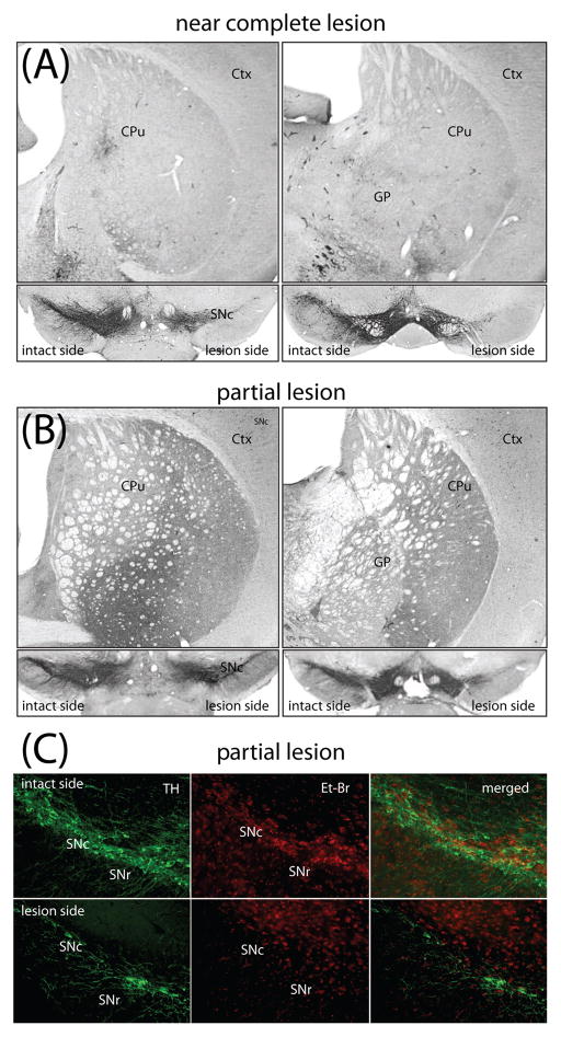
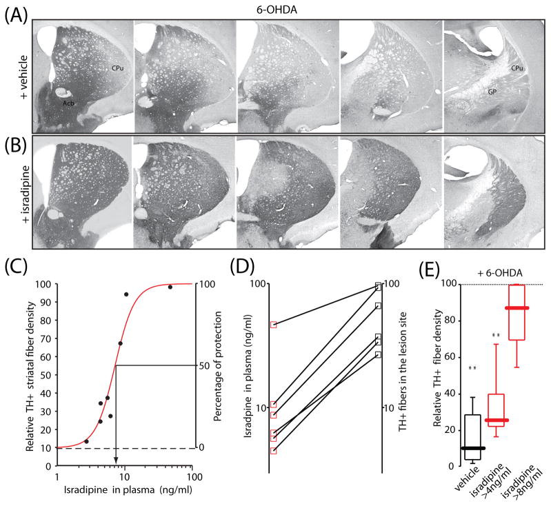
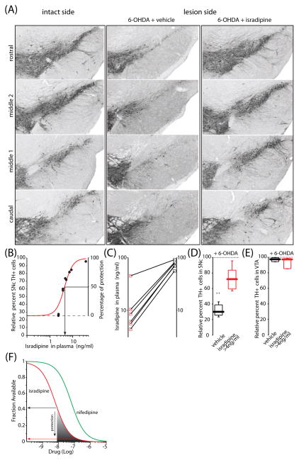
The motor symptoms of Parkinson's disease (PD) are due to the progressive loss of dopamine (DA) neurons in substantia nigra pars compacta (SNc). Nothing is known to slow the progression of the disease, making the identification of potential neuroprotective agents of great clinical importance. Previous studies using the 1-methyl-4-phenyl-1,2,3,6-tetrahydropyridine (MPTP) model of PD have shown that antagonism of L-type Ca2+ channels protects SNc DA neurons. However, this was not true in a 6-hydroxydopamine (6-OHDA) model. One potential explanation for this discrepancy is that protection in the 6-OHDA model requires greater antagonism of Cav1.3 L-type Ca2+ channels thought to underlie vulnerability and this was not achievable with the low affinity dihydropyridine (DHP) antagonist used. To test this hypothesis, the DHP with the highest affinity for Cav1.3L-type channels-isradipine-was systemically administered and then the DA toxin 6-OHDA injected intrastriatally. Twenty-five days later, neuroprotection and plasma concentration of isradipine were determined. This analysis revealed that isradipine produced a dose-dependent sparing of DA fibers and cell bodies at concentrations achievable in humans, suggesting that isradipine is a potentially viable neuroprotective agent for PD.
Copyright © 2011 Elsevier Inc. All rights reserved.
Figures
Similar articles
-
Lower Affinity of Isradipine for L-Type Ca2+ Channels during Substantia Nigra Dopamine Neuron-Like Activity: Implications for Neuroprotection in Parkinson's Disease.J Neurosci. 2017 Jul 12;37(28):6761-6777. doi: 10.1523/JNEUROSCI.2946-16.2017. Epub 2017 Jun 7. J Neurosci. 2017. PMID: 28592699 Free PMC article.
-
Isradipine attenuates MPTP-induced dopamine neuron degeneration by inhibiting up-regulation of L-type calcium channels and iron accumulation in the substantia nigra of mice.Oncotarget. 2017 Jul 18;8(29):47284-47295. doi: 10.18632/oncotarget.17618. Oncotarget. 2017. PMID: 28521299 Free PMC article.
-
The Potential of L-Type Calcium Channels as a Drug Target for Neuroprotective Therapy in Parkinson's Disease.Annu Rev Pharmacol Toxicol. 2019 Jan 6;59:263-289. doi: 10.1146/annurev-pharmtox-010818-021214. Annu Rev Pharmacol Toxicol. 2019. PMID: 30625283 Review.
-
Nimodipine, an L-type calcium channel blocker attenuates mitochondrial dysfunctions to protect against 1-methyl-4-phenyl-1,2,3,6-tetrahydropyridine-induced Parkinsonism in mice.Neurochem Int. 2016 Oct;99:221-232. doi: 10.1016/j.neuint.2016.07.003. Epub 2016 Jul 7. Neurochem Int. 2016. PMID: 27395789
-
Calcium, ageing, and neuronal vulnerability in Parkinson's disease.Lancet Neurol. 2007 Oct;6(10):933-8. doi: 10.1016/S1474-4422(07)70246-6. Lancet Neurol. 2007. PMID: 17884683 Review.
Cited by
-
DNA variants in CACNA1C modify Parkinson disease risk only when vitamin D level is deficient.Neurol Genet. 2016 Apr 12;2(3):e72. doi: 10.1212/NXG.0000000000000072. eCollection 2016 Jun. Neurol Genet. 2016. PMID: 27123490 Free PMC article.
-
The Role of Calcium and Iron Homeostasis in Parkinson's Disease.Brain Sci. 2024 Jan 17;14(1):88. doi: 10.3390/brainsci14010088. Brain Sci. 2024. PMID: 38248303 Free PMC article. Review.
-
Neuro-Restorative Effect of Nimodipine and Calcitriol in 1-Methyl 4-Phenyl 1,2,3,6 Tetrahydropyridine-Induced Zebrafish Parkinson's Disease Model.J Korean Neurosurg Soc. 2024 Sep;67(5):510-520. doi: 10.3340/jkns.2023.0189. Epub 2023 Dec 22. J Korean Neurosurg Soc. 2024. PMID: 38130142 Free PMC article.
-
L-type Calcium Channels are Involved in Iron-induced Neurotoxicity in Primary Cultured Ventral Mesencephalon Neurons of Rats.Neurosci Bull. 2020 Feb;36(2):165-173. doi: 10.1007/s12264-019-00424-2. Epub 2019 Sep 3. Neurosci Bull. 2020. PMID: 31482520 Free PMC article.
-
Determinants of dopaminergic neuron loss in Parkinson's disease.FEBS J. 2018 Oct;285(19):3657-3668. doi: 10.1111/febs.14607. Epub 2018 Aug 14. FEBS J. 2018. PMID: 30028088 Free PMC article. Review.
References
Publication types
MeSH terms
Substances
Grants and funding
LinkOut - more resources
Full Text Sources
Other Literature Sources
Miscellaneous
