. 2011 Jul;85(13):6795-808.
doi: 10.1128/JVI.02074-10. Epub 2011 Apr 20.

Rhinovirus-induced barrier dysfunction in polarized airway epithelial cells is mediated by NADPH oxidase 1

Affiliations

Rhinovirus-induced barrier dysfunction in polarized airway epithelial cells is mediated by NADPH oxidase 1

Adam T Comstock et al. J Virol. 2011 Jul.

Abstract

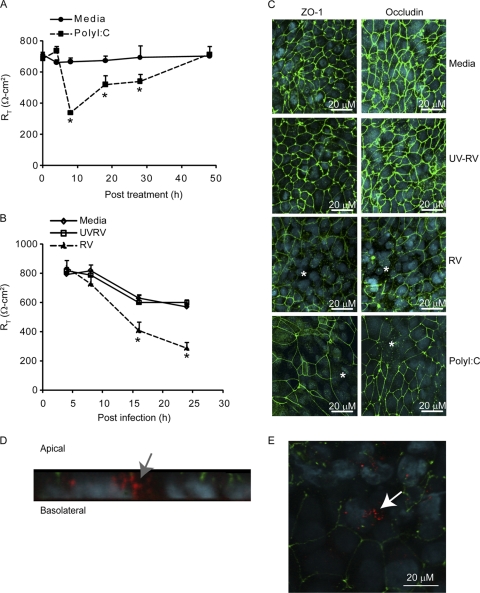
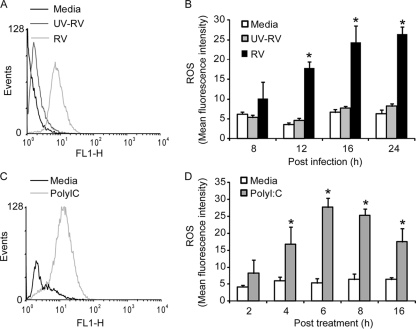
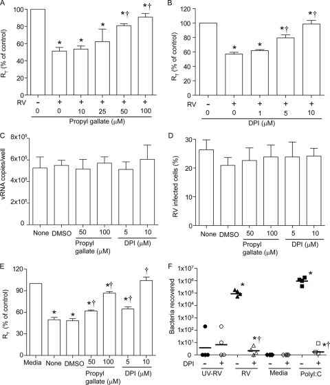
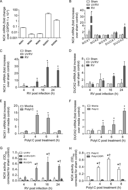
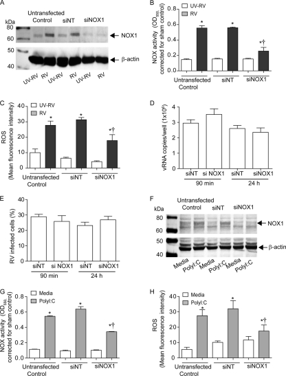
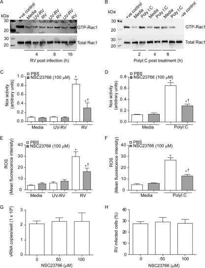
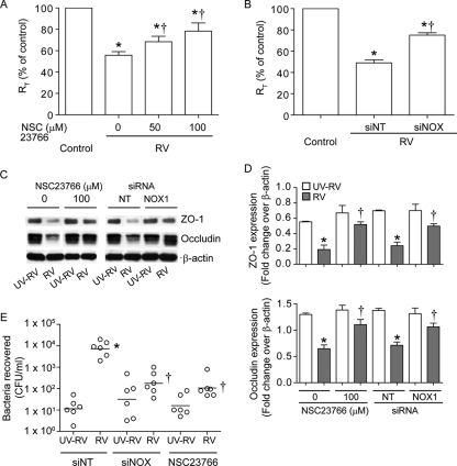
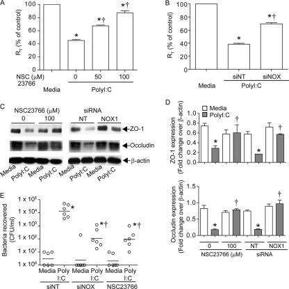
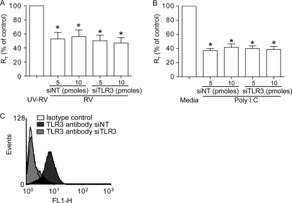
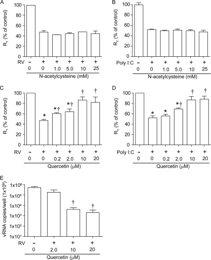
Previously, we showed that rhinovirus (RV), which is responsible for the majority of common colds, disrupts airway epithelial barrier function, as evidenced by reduced transepithelial resistance (R(T)), dissociation of zona occludins 1 (ZO-1) from the tight junction complex, and bacterial transmigration across polarized cells. We also showed that RV replication is required for barrier function disruption. However, the underlying biochemical mechanisms are not known. In the present study, we found that a double-stranded RNA (dsRNA) mimetic, poly(I:C), induced tight junction breakdown and facilitated bacterial transmigration across polarized airway epithelial cells, similar to the case with RV. We also found that RV and poly(I:C) each stimulated Rac1 activation, reactive oxygen species (ROS) generation, and Rac1-dependent NADPH oxidase 1 (NOX1) activity. Inhibitors of Rac1 (NSC23766), NOX (diphenylene iodonium), and NOX1 (small interfering RNA [siRNA]) each blocked the disruptive effects of RV and poly(I:C) on R(T), as well as the dissociation of ZO-1 and occludin from the tight junction complex. Finally, we found that Toll-like receptor 3 (TLR3) is not required for either poly(I:C)- or RV-induced reductions in R(T). Based on these results, we concluded that Rac1-dependent NOX1 activity is required for RV- or poly(I:C)-induced ROS generation, which in turn disrupts the barrier function of polarized airway epithelia. Furthermore, these data suggest that dsRNA generated during RV replication is sufficient to disrupt barrier function.


Figures

Fig. 1.
Fig. 1.
Poly(I:C) decreases RT by dissociating ZO-1 and occludin from tight junction complexes, similar to RV. Polarized airway epithelial cells grown in Transwells were treated with 500 ng/ml poly(I:C) and incubated for up to 48 h (A) or were infected apically with RV or UV-RV for 90 min, after which the infection medium was replaced with fresh medium and the cells were incubated for up to 24 h (B). RT was measured at various time points. Results represent means ± standard errors of the means (SEM) calculated for three independent experiments carried out in triplicate. *, different from medium control or UV-RV- or sham-treated cells (P ≤ 0.05 by ANOVA). In some experiments, cells were treated with poly(I:C) (C), infected with RV or UV-RV (C), or treated with rhodamine-labeled poly(I:C) (D and E) as described above, fixed in methanol, and immunostained with antibody to ZO-1 or occludin (green). Nuclei were stained with DAPI (4′,6-diamidino-2-phenylindole) (blue). Asterisks in panel C represent dissociation of ZO-1 or occludin from the periphery of cells. Arrows in panels D and E represent rhodamine-labeled poly(I:C) below the apical surface and within the cells, respectively. Images are representative of three independent experiments.
Fig. 2.
Fig. 2.
RV infection or poly(I:C) treatment stimulates ROS generation in polarized airway epithelial cells. Polarized 16HBE14o− cells were infected with RV or UV-RV (A and B) or treated with poly(I:C) (C and D). Generation of ROS was measured by flow cytometry using carboxy-H2DCFDA 24 h after RV infection (A) or 8 h after poly(I:C) treatment (C). The kinetics of ROS generation was measured in RV-infected (B) or poly(I:C)-treated (D) cells by flow cytometry. Histograms are representative of four independent experiments. Data in panels B and D are means ± SEM for four independent experiments carried out in duplicate. *, different from medium control or UV-RV-treated cells (P ≤ 0.05 by ANOVA).
Fig. 3.
Fig. 3.
Propyl gallate and DPI inhibit RV- or poly(I:C)-induced barrier disruption in polarized airway epithelial cells. Polarized 16HBE14o− cells were infected with RV or UV-RV for 90 min, and the infection medium was replaced with medium containing propyl gallate (A) or DPI (B). RT was measured after 24 h and expressed as a percentage relative to UV-RV-treated controls. (C) RNAs were extracted from selected groups of RV-infected cells at 24 h postinfection, and the number of vRNA copies was determined. (D) Percentage of cells infected with RV, assessed by flow cytometry. (E) For poly(I:C)-treated cells, propyl gallate and DPI were added 30 min after adding poly(I:C), and RT was measured after 8 h and expressed as a percentage relative to similarly treated medium controls. Data represent means ± SEM. (F) Polarized cells were treated with RV, UV-RV, or poly(I:C) as described above in the presence or absence of 10 μM DPI for 8 h [for poly(I:C)-treated cells] or 24 h (for RV-infected cells), NTHI was added to the apical chamber, and the number of bacteria in the basolateral medium after 3 h was determined by plating. Data represent ranges and medians. *, different from respective UV-RV- or medium-treated controls (P ≤ 0.05); †, different from RV-infected or poly(I:C)-treated cells in the absence of propyl gallate or DPI (P ≤ 0.05 by ANOVA [A to E] or ANOVA on ranks [F]). DMSO, dimethyl sulfoxide.
Fig. 4.
Fig. 4.
RV and poly(I:C) increase mRNA expression of NOX1 and DUOX2 and enhance NOX activity. Total RNAs were isolated from medium-treated cells (A) and from sham-, RV-, and UV-RV-infected cells (B), and expression of NOX1, NOX2, NOX5, DUOX1, and DUOX2 was measured by qPCR. The kinetics of NOX1 and DUOX2 mRNA expression was determined for RV-infected cells (C and D) and poly(I:C)-treated cells (E and F). The kinetics of NOX enzymatic activity was measured at various time points after RV infection (G) or poly(I:C) treatment (H) by cytochrome c reduction assay in the presence or absence of DPI. Data represent means ± SEM calculated for 3 or 4 independent experiments carried out in duplicate. *, different from sham- or medium-treated controls (P ≤ 0.05); †, different from NOX enzymatic activity determined in the presence of DPI (P ≤ 0.05 by ANOVA).
Fig. 5.
Fig. 5.
RV- or poly(I:C)-induced NOX1 expression contributes to increased total NOX activity and ROS generation. 16HBE14o− cells were transfected with siNT or siNOX1. Two days later, cells were infected with RV or treated with poly(I:C) as described in the text. Cells were harvested 16 h (for RV-infected cells) or 8 h [for poly(I:C)-treated cells] after treatment and assessed for expression of NOX1 by Western blot analysis (A and F), for total NOX activity by measuring cytochrome c reduction (B and G), and for ROS generation by flow cytometry using carboxy-H2DCFDA (C and H). The vRNA copy number (D) and % of RV-infected cells (E) were determined for siNT- and siNOX1-transfected cells 24 h after RV infection. Data represent means ± SEM calculated for 3 or 4 independent experiments carried out in duplicate. *, different from respective controls (P ≤ 0.05); †, different from respective siNT-treated controls (P ≤ 0.05 by ANOVA).
Fig. 6.
Fig. 6.
RV or poly(I:C)-induced Rac1 activity partially contributes to total NOX activity and ROS generation. 16HBE14o− cells were infected with RV (A) or treated with poly(I:C) (B), and Rac1 activity was determined by pulldown assay at various time points. Medium- and UV-RV-treated cells were used as controls. +ve control and −ve control, cell lysates from medium-treated cells incubated with GTPγS and GDP, respectively. Images are representative of 3 experiments. Cells were then infected with RV or UV-RV (C and E) or treated with medium or poly(I:C) (D and F) and incubated in the presence or absence of NSC27366. Cells were harvested after 8 h [for poly(I:C)-treated cells] or 24 h (for RV-infected cells) and then examined for NOX activity (C and D) and ROS generation (E and F). The vRNA copy number (G) and % of RV-infected cells (H) were determined for siNT- and siNOX-transfected cells 24 h after RV infection. Data represent means ± SEM calculated for 3 or 4 independent experiments carried out in duplicate. *, different from respective controls (P ≤ 0.05); †, different from RV-infected or poly(I:C)-treated cells in the absence of NSC23766 (P ≤ 0.05 by ANOVA).
Fig. 7.
Fig. 7.
Rac1 and NOX1 are required for RV-induced disruption of barrier function. (A) Polarized airway epithelial cells grown in Transwells were infected with RV or UV-RV as described in the text and incubated for 24 h in the presence or absence of various concentrations of NSC23766. RT was expressed as a percentage relative to UV-RV-infected controls. (B) Cells growing in Transwells were transfected with NT or NOX1 siRNA, and 2 days later, cells were infected with RV or UV-RV. RT was measured after 24 h and expressed as a percentage relative to the respective UV-RV controls. (C) The cytoskeleton fraction was subjected to Western blot analysis with antibody to ZO-1 or occludin, and the image is representative of 3 independent experiments. (D) Quantification of the ratio of occludin or ZO-1 to β-actin. (E) Twenty-four hours after RV or UV-RV infection, nontypeable H. influenzae was added to the apical chamber and bacteria in the basolateral chamber were quantified to assess bacterial transmigration across polarized airway epithelial cells. Data represent means ± SEM (A, B, and D) or ranges with medians (E) calculated for 3 or 4 independent experiments carried out in duplicate. *, different from respective controls (P ≤ 0.05); †, different from RV-infected cells in the absence of NSC23766 or from NT siRNA-transfected cells infected with RV (P ≤ 0.05 by ANOVA or ANOVA on ranks).
Fig. 8.
Fig. 8.
Poly(I:C)-induced barrier disruption requires both Rac1 and NOX1. (A) Polarized epithelial cells were treated with medium or poly(I:C) for 8 h in the presence or absence of NSC23766, and RT was measured. (B) Cells transfected with NT or NOX1 siRNA were treated with poly(I:C), and RT was measured after 8 h. RT is expressed as a percentage relative to the respective medium controls. (C and D) ZO-1 and occludin protein levels in the cytoskeleton fraction were determined by Western blot analysis followed by densitometry and are expressed as the ratio of ZO-1 or occludin to β-actin. The Western blot is representative of 3 experiments. (E) Eight hours after poly(I:C) treatment, bacterial transmigration across polarized airway epithelial cells was measured as described in the text. Data represent means ± SEM (A, B, and D) or ranges with medians (E) calculated for 3 or 4 independent experiments carried out in duplicate. *, different from respective controls (P ≤ 0.05); †, different from RV-infected cells in the absence of NSC23766 or from NT siRNA-transfected cells infected with RV (P ≤ 0.05 by ANOVA or ANOVA on ranks).
Fig. 9.
Fig. 9.
TLR3 is not required for either RV- or poly(I:C)-induced reduction in RT. Cells transfected with NT or TLR3 siRNA were treated with RV (A) or poly(I:C) (B), and RT was measured after 24 or 8 h, respectively. Data are expressed as percentages relative to the respective UV-RV- or medium-treated controls and represent means ± SEM calculated for 3 or 4 independent experiments carried out in duplicate. *, different from respective controls (P ≤ 0.05 by ANOVA). (C) Expression of TLR3 in siNT- or siTLR3-transfected cells was determined by flow cytometry. The histogram shown is representative of 3 experiments.
Fig. 10.
Fig. 10.
Quercetin, but not N-acetylcysteine, blocks disruptive effects of RV and poly(I:C) on RT. Polarized 16HBE14o− cells were infected with RV or UV-RV for 90 min, the infection medium was replaced with medium containing N-acetylcysteine (A) or quercetin (C), and RT was measured 24 h later. Cells were also treated with poly(I:C) in the presence of N-acetylcysteine (B) or quercetin (D), and RT was measured after 8 h. Data are expressed as percentages relative to medium- or UV-RV-treated controls, as appropriate. (E) Total RNA was isolated from cells infected with RV in the presence of quercetin, and the number of copies of vRNA was determined by qPCR. Data represent means ± SEM calculated for 2 or 3 independent experiments carried out in duplicate. *, different from respective UV-RV- or medium-treated controls (P ≤ 0.05); †, different from RV-infected or poly(I:C)-treated cells in the absence of quercetin (P ≤ 0.05 by ANOVA).

Similar articles

Cited by

References

    1. Arruda E., Pitkaranta A., Witek T. J., Jr., Doyle C. A., Hayden F. G. 1997. Frequency and natural history of rhinovirus infections in adults during autumn. J. Clin. Microbiol. 35:2864–2868 - PMC - PubMed
    1. Avadhanula V., et al. 2006. Respiratory viruses augment the adhesion of bacterial pathogens to respiratory epithelium in a viral species- and cell type-dependent manner. J. Virol. 80:1629–1636 - PMC - PubMed
    1. Bedard K., Krause K. H. 2007. The NOX family of ROS-generating NADPH oxidases: physiology and pathophysiology. Physiol. Rev. 87:245–313 - PubMed
    1. Berube J., Bourdon C., Yao Y., Rousseau S. 2009. Distinct intracellular signaling pathways control the synthesis of IL-8 and RANTES in TLR1/TLR2, TLR3 or NOD1 activated human airway epithelial cells. Cell. Signal. 21:448–456 - PubMed
    1. Biagioli M. C., Kaul P., Singh I., Turner R. B. 1999. The role of oxidative stress in rhinovirus induced elaboration of IL-8 by respiratory epithelial cells. Free Radic. Biol. Med. 26:454–462 - PubMed

Publication types

MeSH terms