. 2011 Apr 13;31(15):5744-54.
doi: 10.1523/JNEUROSCI.6810-10.2011.

Reduced BACE1 activity enhances clearance of myelin debris and regeneration of axons in the injured peripheral nervous system

Affiliations

Reduced BACE1 activity enhances clearance of myelin debris and regeneration of axons in the injured peripheral nervous system

Mohamed H Farah et al. J Neurosci. .

Abstract

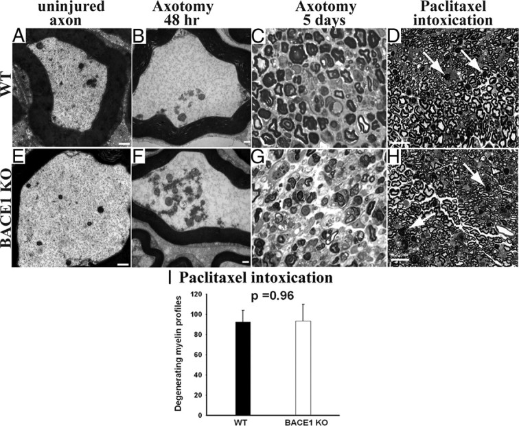
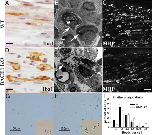
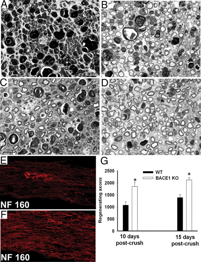
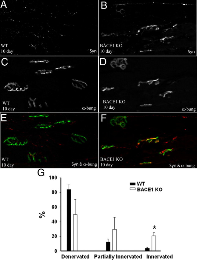
β-Site amyloid precursor protein (APP) cleaving enzyme 1 (BACE1) is an aspartyl protease best known for its role in generating the amyloid-β peptides that are present in plaques of Alzheimer's disease. BACE1 has been an attractive target for drug development. In cultured embryonic neurons, BACE1-cleaved N-terminal APP is further processed to generate a fragment that can trigger axonal degeneration, suggesting a vital role for BACE1 in axonal health. In addition, BACE1 cleaves neuregulin 1 type III, a protein critical for myelination of peripheral axons by Schwann cells during development. Here, we asked whether axonal degeneration or axonal regeneration in adult nerves might be affected by inhibition or elimination of BACE1. We report that BACE1 knock-out and wild-type nerves degenerated at a similar rate after axotomy and to a similar extent in the experimental neuropathies produced by administration of paclitaxel and acrylamide. These data indicate N-APP is not the sole culprit in axonal degeneration in adult nerves. Unexpectedly, however, we observed that BACE1 knock-out mice had markedly enhanced clearance of axonal and myelin debris from degenerated fibers, accelerated axonal regeneration, and earlier reinnervation of neuromuscular junctions, compared with littermate controls. These observations were reproduced in part by pharmacological inhibition of BACE1. These data suggest BACE1 inhibition as a therapeutic approach to accelerate regeneration and recovery after peripheral nerve damage.


Figures

Figure 1.
Figure 1.
Similar degree of axonal and myelin degeneration in WT and BACE1 KO sciatic nerves after axotomy-induced Wallerian degeneration and intoxication with paclitaxel. A, B, E, and F are electron micrographs, and C, D, G, and H are semithin (1 μm) plastic sections stained with toluidine blue. A, Uninjured WT axon with normal axoplasm. B, WT at 48 h after crush showing conversion of the axonal cytoskeleton to granular debris. C, WT nerve at 5 d after crush showing degeneration of myelin. D, WT nerve intoxicated with paclitaxel. The arrows point to degenerating myelinated fibers. E, Uninjured BACE1 KO axon with normal axoplasm. F, BACE1 KO at 48 h after crush showing conversion of the axonal cytoskeleton to granular debris, similar to WT in B. G, BACE1 KO nerve at 5 d after crush showing degeneration of myelin. H, BACE1 KO nerve intoxicated with paclitaxel. The arrows point to degenerating myelinated fibers. I, Quantification of degenerating myelinated fibers in the whole cross-sectional area of nerves of mice intoxicated with paclitaxel. N = 3 per genotype. Values are mean ± SEM. Scale bars: A, B, D, F, 500 nm; C, F, G, H, 100 μm.
Figure 2.
Figure 2.
Enhanced clearance of axonal debris and enhanced axonal regeneration in YFP-BACE1 KO nerves compared with YFP-WT nerves after axotomy. These images are projections of Z-stacks from whole-mounted nerves to show the YPF-positive axonal debris and YFP-positive regenerated axons through the depth of the nerves. A, YFP-WT nerve at 7 d after transection. Fragmented YFP debris (arrowhead) is present. B, YFP-BACE1 KO nerve at 7 d after transection. YFP debris (arrowhead) is mostly cleared from the nerve. C, YFP-WT nerve at 10 d after crush. Fragmented YFP debris (arrowhead) is present at a segment 10 mm distal to the crush and few regenerated YFP-positive axons (arrow) are growing beneath the debris. D, YFP-BACE1 KO nerve at 10 d after crush. Numerous regenerated YFP-positive axons are present at segment 10 mm distal to crush site and (arrows). There are scanty clumps of YFP-positive axonal debris remaining. Scale bars, 100 μm. Laser-scanning intensity was increased in C and D to reveal small-diameter YFP-positive regenerated axons.
Figure 3.
Figure 3.
Enhanced phagocytosis by BACE1 KO macrophages both in vivo and in vitro. A–F are micrographs of distal stumps at 5 d after injury. A and D are stained for Iba1 (ionized calcium-binding adaptor molecule 1), a marker for macrophages. B and E are electron micrographs. C and F are stained for MBP. G and H are bright-field images of cultured peritoneal macrophages. A, The majority of Iba1-positive macrophages in WT nerve are elongated at 5 d after cut. B, Undigested myelin debris and ovoids are present in WT nerve at 5 d after crush. C, WT nerve with abundant elongated MBP-positive myelin debris at 5 d after cut. D, Iba1-positive BACE1 KO macrophages are larger in size and appear activated at 5 d after cut. E, Activated macrophages filled with lipid droplets (arrow) are present in BACE1 KO nerves at 5 d after crush. F, BACE1 KO nerve with broken down MBP-positive myelin debris at 5 d after cut. G, WT macrophages after incubation with IgG-coated beads for 5 min. H, BACE1 KO macrophages after incubation with IgG-coated beads for 5 min. I, Quantification of IgG-coated beads per individual cells after 5 min of phagocytosis. Data are from three independent experiments. Values are mean ± SEM. Scale bars: A, D, 10 μm; B, E, 2 μm; C, F, 20 μm; G, H, 100 μm.
Figure 4.
Figure 4.
Accelerated rate of axonal outgrowth in BACE1 KO versus WT after crush injury. A and B are longitudinally sectioned sciatic nerves stained for GAP43 at 3 d after crush. In 2 mm segment distal to the crush, BACE1 KO nerve (B) has more regenerating axons that reach further than those axons of WT littermate nerve (A). C and D are longitudinal sections of nerves filled with Neurobiotin to anterogradely label regenerating axons at 5 d after crush. BACE1 KO nerve (D) has significantly more regenerated axons that reach further distally compared with WT littermate nerve (C). Scale bars: A, B, 100 μm; C, D, 500 μm. The stars in C and D indicate approximate sites of crush.
Figure 5.
Figure 5.
Extensive axonal regeneration and large polyaxonal pockets in the distal segment of crushed BACE1 KO sciatic nerve at 5 d after crush. A–F are electron micrographs. A, WT nerve. Abundant myelin debris and ovoids and few regenerating sprouts are present (arrow). B, BACE1 KO nerve. Myelin debris is cleared and numerous regenerating sprouts (arrows) are present. C, WT nerve. Few polyaxonal pockets are present. D, BACE1 KO nerve. Numerous polyaxonal pockets (arrows) are present. E, WT nerve. A degenerating myelinated fiber has one (arrow) sprout growing beneath the basal lamina membrane. F, BACE1 KO nerve. Multiple sprouts (arrows) are growing within individual degenerating fibers. G, The BACE1 KO fibers exhibit enhanced number of regenerating sprouts within a single basal lamina membrane compared with WT fibers. For example, 66% of the BACE1 KO fibers have two or more regenerating sprouts compared with 31% of the WT fibers, which have two or more sprouts. Scale bars: A–D, 2 μm.
Figure 6.
Figure 6.
BACE1 KO regenerating axons form a central channel of microtubule cluster; WT axons do not. A, Regenerated axon with normally distributed cytoskeleton structures such as microtubules and neurofilament. B, Regenerated BACE1 KO axon with a central channel of microtubules.
Figure 7.
Figure 7.
Significantly more regenerated axons at 10 and 15 d after crush in the distal segments of crushed BACE1 KO versus WT sciatic nerve. A–D are semithin (1 μm) plastic sections stained with toluidine blue, and E and F are frozen sections stained for phosphorylated neurofilament (NF 160). A, In WT nerve, a large amount of myelin debris in various stages of degradation is present at 10 d after crush with some regenerating axons. B, In BACE1 KO nerve, large number of regenerating axons, some foamy macrophages, and myelin debris are present at 10 d after crush. C, In WT nerve, regenerating axons are present at 15 d after crush. D, In BACE1 KO nerve, significantly more regenerating axons are present at 15 d after crush (compare C, D). E, WT nerve segment at 10–12 mm distal to the crush site. A few NF 160-positive axons are regenerating. F, BACE1 KO nerve segment at 10–12 mm distal to the crush site. Significant numbers of NF 160-positive axons are regenerating. G, Axon counts at 10 and 15 d after crush. At both time points, BACE1 KO nerves have significantly more regenerated axons than WT nerves. *p < 0.05. n = 3 nerves per genotype at each time point. Values are mean ± SEM. Scale bar: A–D, 10 μm.
Figure 8.
Figure 8.
Robust reinnervation of neuromuscular junctions in BACE1 KO versus WT at 10 d after crush. Ten days after nerve crush, the gastrocnemius muscle was harvested from BACE1 KO and WT mice. A and B are stained for synaptophysin; C and D are stained for α-bungarotoxin. E and F are merged images of A and C, and B and D, respectively. Reinnervating presynaptic endings at neuromuscular junctions in BACE1 KO appeared more mature than those in WT. G, Counts of neuromuscular junctions in WT and BACE1 KO mice. BACE1 KO muscles have significantly more reinnervated junctions. N = 3 per genotype. Values are mean ± SEM. *p < 0.05.
Figure 9.
Figure 9.
Two structurally distinct small molecule BACE1 inhibitors accelerate debris clearance and axonal regeneration after axotomy in WT mice. A, YFP-WT nerve at 7 d after crush. Fragmented YFP debris (arrowheads) is present. Few regenerated YFP-positive axons (arrows) are growing beneath the debris. B, YFP-WT nerve treated with BACE1 inhibitor IV for 7 d after nerve crush. YFP debris (arrowheads) is more fragmented in the nerve. Numerous regenerated YFP-positive axons (arrows) are growing beneath the debris. C, WT nerve at 7 d after crush. A few regenerating axons (arrows) are present. D, WT nerve treated with BACE1 inhibitor IV at 7 d after crush. The nerve has more regenerated axons (arrows) than WT littermate nerve in C. More post-phagocytic macrophages (arrowhead) are present. E, WT foot stained for β-tubulin III to reveal the intraplantar nerves. F, WT foot stained for β-tubulin III 2 weeks after sciatic nerve crush. Regenerated axons have not arrived at the foot yet. G, WT foot stained for β-tubulin III 2 weeks after sciatic nerve crush and treatment with BACE1 inhibitor WAY 258131. β-Tubulin III-positive fibers are regenerated axons in the intraplantar nerves. A and B images are projections of Z-stacks from whole-mounted nerves to show the YFP-positive axonal debris through the depth of the nerve. C and D are electron micrographs. Scale bars: A, B, 100 μm; C, D, 2 μm.
Figure 10.
Figure 10.
Accelerated axonal regeneration of BACE1 KO axons. A, Above is a schematic of a reciprocal sciatic nerve transplantation between BACE1 KO and WT. Below is a representative image of a nerve that was transplanted using this nerve graft technique. The arrows point to where the transplanted segment was sutured in. B, WT nerve segment transplanted into a BACE1 KO host nerve. BACE1 KO axons regenerated through the WT graft well. The arrows point to regenerated BACE1 KO axons. C, BACE1 nerve segment transplanted into a wild-type host. A few WT axons grow through BACE1 graft (arrow). Scale bars: B, C, 10 μm. D, BACE1 KO DRG explants had axons 119.0 and 150.0% longer on average than those of WT DRG explants at 2 and 4 d after culture. BACE1 inhibitor IV (50 nm) increased axonal outgrowth of WT DRG explants by 98.0% at 4 d after culture. Values are mean ± SEM. *p < 0.001.

Similar articles

Cited by

References

    1. Araki W, Kitaguchi N, Tokushima Y, Ishii K, Aratake H, Shimohama S, Nakamura S, Kimura J. Trophic effect of β-amyloid precursor protein on cerebral cortical neurons in culture. Biochem Biophys Res Commun. 1991;181:265–271. - PubMed
    1. Barrette B, Hébert MA, Filali M, Lafortune K, Vallières N, Gowing G, Julien JP, Lacroix S. Requirement of myeloid cells for axon regeneration. J Neurosci. 2008;28:9363–9376. - PMC - PubMed
    1. Beirowski B, Berek L, Adalbert R, Wagner D, Grumme DS, Addicks K, Ribchester RR, Coleman MP. Quantitative and qualitative analysis of Wallerian degeneration using restricted axonal labelling in YFP-H mice. J Neurosci Methods. 2004;134:23–35. - PubMed
    1. Beirowski B, Adalbert R, Wagner D, Grumme DS, Addicks K, Ribchester RR, Coleman MP. The progressive nature of Wallerian degeneration in wild-type and slow Wallerian degeneration (WldS) nerves. BMC Neurosci. 2005;6:6. - PMC - PubMed
    1. Boivin A, Pineau I, Barrette B, Filali M, Vallières N, Rivest S, Lacroix S. Toll-like receptor signaling is critical for Wallerian degeneration and functional recovery after peripheral nerve injury. J Neurosci. 2007;27:12565–12576. - PMC - PubMed

MeSH terms

LinkOut - more resources