. 2011 Apr 12;19(4):470-83.
doi: 10.1016/j.ccr.2011.02.007.

Deletion of p120-catenin results in a tumor microenvironment with inflammation and cancer that establishes it as a tumor suppressor gene

Affiliations

Deletion of p120-catenin results in a tumor microenvironment with inflammation and cancer that establishes it as a tumor suppressor gene

Douglas B Stairs et al. Cancer Cell. .

Abstract

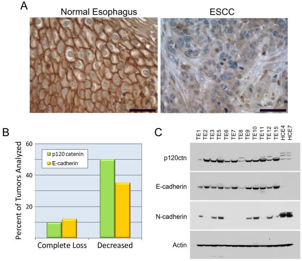
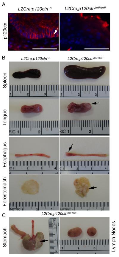
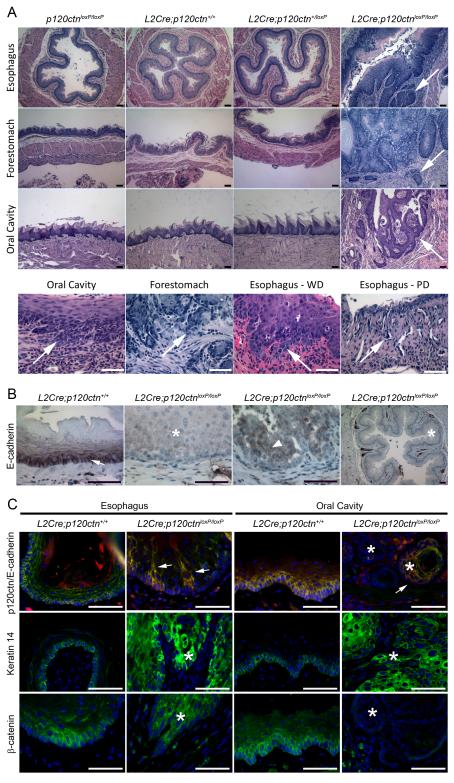
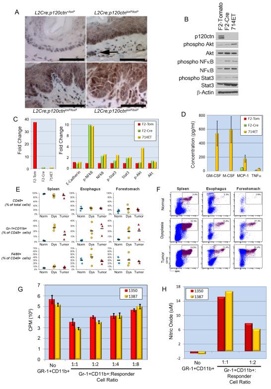
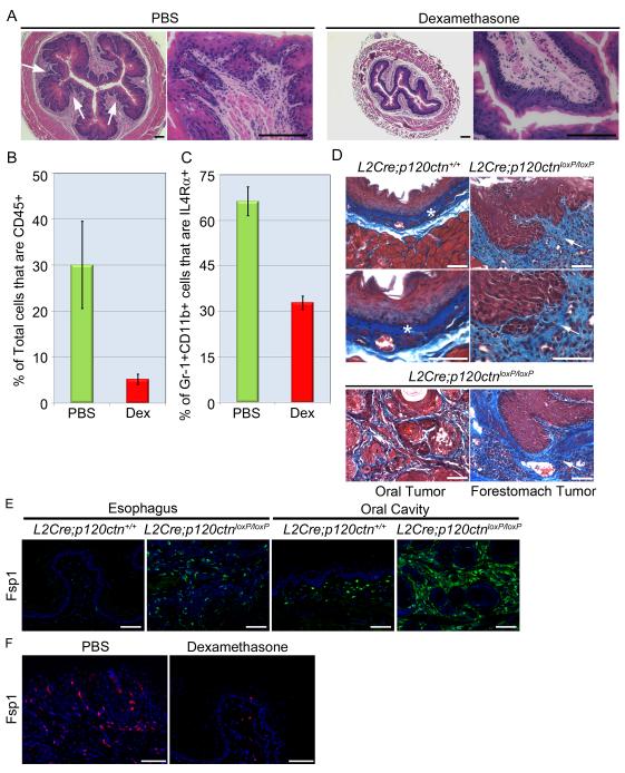
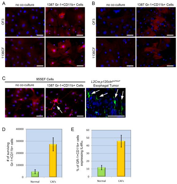
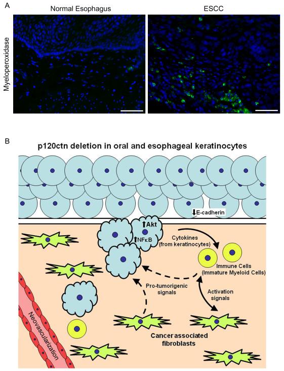
p120-catenin (p120ctn) interacts with E-cadherin, but to our knowledge, no formal proof that p120ctn functions as a bona fide tumor suppressor gene has emerged to date. We report herein that p120ctn loss leads to tumor development in mice. We have generated a conditional knockout model of p120ctn whereby mice develop preneoplastic and neoplastic lesions in the oral cavity, esophagus, and squamous forestomach. Tumor-derived cells secrete granulocyte macrophage colony-stimulating factor (GM-CSF), macrophage colony-stimulating factor (M-CSF), monocyte chemotactic protein-1 (MCP-1), and tumor necrosis factor-α (TNFα). The tumors contain significant desmoplasia and immune cell infiltration. Immature myeloid cells comprise a significant percentage of the immune cells present and likely participate in fostering a favorable tumor microenvironment, including the activation of fibroblasts.


Figures

Figure 1
Figure 1. P120ctn and E-cadherin expression in ESCC
(A) P120ctn expression by immunohistochemistry (IHC) reveals cell membrane localization in normal tissue, whereas cytoplasmic mislocalization and decreased or absent expression is detected in ESCC. Scale bars=50uM. (B) Quantification of p120ctn and E cadherin expression in ESCC relative to matched adjacent normal esophagus (n=69 paired samples). Fisher’s exact analysis demonstrates that p120ctn and E-cadherin expression are coordinately regulated (p=0.017). (C) Western blot analysis of human esophageal cancer cell lines for p120ctn, E-cadherin, and N-cadherin expression, with ß-actin as loading control.
Figure 2
Figure 2. Gross pathology of L2Cre;p120ctnloxP/loxP mice
(A) Two-month old mice were analyzed by immunofluorescence (IF) for p120ctn expression. Arrow indicates normal cell membrane localization in L2Cre;p120ctn+/+ esophagi (n=4). P120ctn loss is evident in the esophagi of L2Cre;p120ctnloxP/loxP mice (n=4). Non-specific staining of keratins can be observed in the cornified layers. Scale bars=50uM. (B) Gross lesions were detected in the spleen, tongue, esophagus and squamous forestomach of 9-12 month old L2Cre;p120ctnloxP/loxP mice. A reference ruler (centimeters) is included in each image for size comparison. Arrows indicate the location of tumors. (C) Gross pathology of a squamous forestomach tumor (indicated by an arrow) and two enlarged lymph nodes (normally approximately 1-2 mm in diameter) from a L2Cre;p120ctnloxP/loxP mouse. See also Table S1 and Figure S1. A total of 23 L2Cre;p120ctnloxP/loxP mice and 24 control (p120ctnloxP/loxP− n=5, L2Cre;p120ctn+/+ − n=10 and L2Cre;p120ctn+/loxP − n=8) mice were evaluated.
Figure 3
Figure 3. Histologic analysis of tumors from L2Cre;p120ctnloxP/loxP mice
(A) H&E analyses of the esophagi, forestomachs and oral cavities of 12 month old L2Cre;p120ctnloxP/loxP mice (n=17) revealed invasive squamous cell cancers. All control mice (p120ctnloxP/loxP− n=5, L2Cre;p120ctn+/+ − n=10 and L2Cre;p120ctn+/loxP − n=8) had no lesions. The bottom row of images denotes higher magnification views of L2Cre;p120ctnloxP/loxP mice containing regions of invasive cancer (denoted by arrows). WD=well differentiated tumor, PD=poorly differentiated tumor. Scale bars=50uM. (B) IHC staining of 12 month old mice for E-cadherin expression (n=4 for each genotype). Arrow indicates normal membrane localization. Asterisks indicate loss of E-cadherin expression and arrowhead indicates cytoplasmic localization of residual E-cadherin expression. Scale bars=50uM. (C) IF of the esophagi and oral cavities of L2Cre;p120ctn+/+ and L2Cre;p120ctnloxP/loxP mice (n=4 for each) for p120ctn (red)-E-cadherin (green) co-staining, Keratin 14 staining, and β-catenin staining. Arrows denote occasional regions of p120ctn and E-cadherin co-localization. Keratin 14 expression was detected in invading cells of both cancer types. β-catenin was retained but mislocalized to the cytoplasm in esophageal tumors. By contrast, β -catenin expression was lost in oral cavity tumors. Asterisks denote regions of invasive cancers. Scale bars=50uM. See also Figure S2.
Figure 4
Figure 4. Immature myeloid cells are recruited to the esophagi and squamous forestomachs of L2Cre;p120ctnloxP/loxP mice
(A) NFκB upregulation in L2Cre;p120ctnloxP/loxP mice. IHC staining for the NFκB p65 subunit revealed significant up-regulation in hyperplastic, dysplastic and invasive cancer lesions in the esophagi of L2Cre;p120ctnloxP/loxP mice (n=6). Arrows denote NFκB nuclear staining in immune cells within the stroma in L2Cre;p120ctnloxP/loxP mice. Interestingly, these cells were found in early hyperplastic lesions. Scale bars=50uM. (B) Western blot analysis of mouse derived cell lines (F2-Tomato, F2-Cre, 714ET-see methods) were used to investigate p120ctn loss as well as Akt, NFκB and Stat3 activation (performed in triplicate). β-actin is used as a loading control. (C) Densitometry of Western blot data from panel B. All data are normalized to β-actin. (D) Cytokine analysis of conditioned media from F2-Tomato, F2-Cre and 714 ET cells (n=3 for each cell line). ELISAs revealed increased GM-CSF (p=0.040), M-CSF (p=0.035), MCP-1 (p=0.026) and TNFα (p=0.005) in 714ET cells and increased MCP-1 in F2-Cre cells (p=1.6×10⁻⁵). Nondetectable ELISA readings were given a value of zero for graphical purposes. (E) Flow cytometry for CD45+ (total leukocytes) cells, Gr-1+ CD11b+ (immature myeloid) cells and F4/80+ macrophages in the spleen (left), esophagus (middle), and forestomach (right) of control mice (L2Cre;p120ctn+/+ − blue diamonds; n = 4), L2Cre;p120ctnloxP/loxP mice with dysplasia (yellow circles; n = 4), and tumor-bearing (TB) L2Cre;p120ctnloxP/loxP mice (red triangles; n = 3). Black horizontal bars are mean percentages for each group. See p-values in Figure S3. (F) Representative flow cytometric results from panel E are depicted. Cells from the spleen (left), esophagus (middle), and forestomach (right) were gated for CD45 expression and then analyzed for Gr-1(x-axis) versus CD11b (y-axis) expression. Numbers represent percentages of Gr-1+ CD11b+ (immature myeloid cells) among CD45+ cells. (G) Proliferation assay for T-cells co-cultured with Gr-1+CD11b+ cells at the indicated cell ratios. Gr-1+CD11b+ cells suppressed T-cell proliferation (n=6). As the ratio of Gr-1+CD11b+ cells relative to T-cells decreased, T-cell proliferation increased (n=2 with 1350, 1387 representing L2Cre;p120ctnloxP/loxP mice). All p-values < 0.05 except 1:8 ratio for mouse 1387 when compared to no Gr-1+CD11b+ cells. (H) Nitric Oxide (NO) in the conditioned media was determined (n=2 L2Cre;p120ctnloxP/loxP mice-1350, 1387). NO levels were increased in a titratable manner. Error bars indicate SEM. See also Table S2 and Figure S3.
Figure 5
Figure 5. The immune response is involved in the tumor microenvironment in L2Cre;p120ctnloxP/loxP mice
(A) Dexamethasone (Dex) or PBS (control) was administered to 8 month old L2Cre;p120ctnloxP/loxP mice (n=5 in each group) for 7 days. Histologic examination of the esophagi revealed hyperplasia without desmoplasia or inflammation in the Dex-treated mice while invasive esophageal squamous cancer with desmoplasia and inflammation was present in PBS-treated mice. Arrows point to regions of invasion. Scale bars=100uM. (B) FACS analysis for CD45+ cells contained within the esophagi from Dex-treated mice (red bars) and PBS control mice (green bars) (n=5 in each group; p=0.01). Dex treatment reduced CD45+ and Gr-1+CD11b+ cells by 6-fold. (C) FACS analysis for IL-4Rα+ cells contained within the Gr-1+CD11b+ population of cells from Dex-treated mice (red bars) and PBS control mice (green bars) (n=5 in each group; p=0.0016). Dex reduced the percentage of Gr-1+ CD11b+ cells that express IL-4Rα by about 50%. (D) Masson’s trichrome staining on esophagi of L2Cre;p120ctnloxP/loxP mice (n=4) and control (L2Cre;p120ctn+/+) mice. Asterisks indicate the fibrillar collagen layer in the control L2Cre;p120ctn+/+ esophagi. Arrows indicate the abundant fibrillar collagen surrounding invasive esophageal tumors. Invasive oral and forestomach tumors in L2Cre;p120ctnloxP/loxP mice are depicted in the bottom row. Arrows indicate the staining surrounding invasive cancers. Scale bars=50uM. (E) Fsp1 IF with increased expression in esophageal and oral cavity tumors from L2Cre;p120ctnloxP/loxP mice (n=4) compared to control L2Cre;p120ctn+/+ mice (n=4) tissues. Scale bar=50uM. (F) Fsp1 IF of esophagi from L2Cre;p120ctnloxP/loxP mice treated with either PBS or Dex from Figure 5A. Fsp1 expression is decreased in Dex-treated mice (n=5) when compared to PBS treated mice (n=5). Scale bars=50uM. See also Figure S4. Error bars indicate SEM.
Figure 6
Figure 6. Crosstalk between immature myeloid cells and fibroblasts in the tumor microenvironment of L2Cre;p120ctnloxP/loxP mice
(A) Fibroblasts from the oral cavity of wild-type mice (designated as OF3) or oral cancer-associated fibroblasts (CAF; designated as 1136CF)) from L2Cre;p120ctnloxP/loxP mice were analyzed in co-culture with Gr-1+CD11b+ cells for Fsp1 expression by IF. Co-cultured fibroblasts had increased Fsp1 expression (n=6). Scale bars=50uM. (B) Co-cultured fibroblasts had increased αSMA expression (n=6). Scale bars=50uM. (C) An esophageal CAF cell line (955EF) had high basal Fsp1 expression when cultured alone. Co-culturing of 955EF cells (n=6) resulted in even higher Fsp1 expression. Note that the highest Fsp1+ 955EF cells have “dendrite-like” projections, which is evident also in esophageal tumors from L2Cre;p120ctnloxP/loxP mice (n=4). Scale bars=50uM. (D) FACS revealed increased survival of Gr-1+CD11b+ cells in co-culture with CAFs (n=4, p=0.021). (E) FACS demonstrated increased IL-4Rα expression in Gr-1+CD11b+ cells in co-culture with CAFs (n=4, p=0.016). Error bars indicate SEM.
Figure 7
Figure 7. Myeloperoxidase expression in human ESCC
(A) Myeloperoxidase (MPO) expression was analyzed by IF in 14 ESCC samples with matched adjacent normal esophageal mucosa. Representative image is depicted with MPO positive cells present in the tumor stroma (right panel). MPO staining was increased in the tumor stroma and correlates with p120ctn loss (p=0.05, Fisher’s exact analysis). Scale bars=50uM. (B) Model of OSCC and ESCC in conditional p120ctn-deleted mice. Solid lines represent cell-cell interactions emerging from experiments in this study. Dashed lines are potential areas of interactions. See also Figure S5.

Comment in

Similar articles

Cited by

References

    1. Anastasiadis PZ, Moon SY, Thoreson MA, Mariner DJ, Crawford HC, Zheng Y, Reynolds AB. Inhibition of RhoA by p120 catenin. Nat Cell Biol. 2000;2:637–644. - PubMed
    1. Andl CD, Mizushima T, Nakagawa H, Oyama K, Harada H, Chruma K, Herlyn M, Rustgi AK. Epidermal growth factor receptor mediates increased cell proliferation, migration, and aggregation in esophageal keratinocytes in vitro and in vivo. J Biol Chem. 2003;278:1824–1830. - PubMed
    1. Andreu P, Johansson M, Affara NI, Pucci F, Tan T, Junankar S, Korets L, Lam J, Tawfik D, DeNardo DG, et al. FcRgamma activation regulates inflammation-associated squamous carcinogenesis. Cancer Cell. 2010;17:121–134. - PMC - PubMed
    1. Bartlett JD, Dobeck JM, Tye CE, Perez-Moreno M, Stokes N, Reynolds AB, Fuchs E, Skobe Z. Targeted p120-catenin ablation disrupts dental enamel development. PLoS One. 2010;5 - PMC - PubMed
    1. Bass AJ, Watanabe H, Mermel CH, Yu S, Perner S, Verhaak RG, Kim SY, Wardwell L, Tamayo P, Gat-Viks I, et al. SOX2 is an amplified lineage-survival oncogene in lung and esophageal squamous cell carcinomas. Nat Genet. 2009;41:1238–1242. - PMC - PubMed

Publication types

MeSH terms

Associated data