

Age-related changes in the function of autophagy in rat kidneys
- PMID: 21455601
- PMCID: PMC3312632
- DOI: 10.1007/s11357-011-9237-1
Age-related changes in the function of autophagy in rat kidneys
Abstract
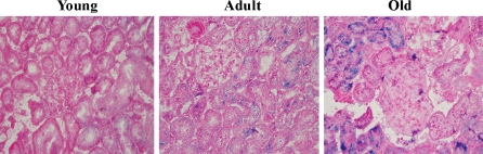
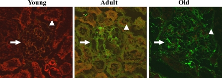
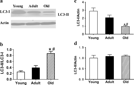
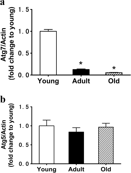
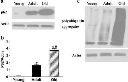
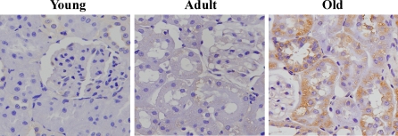
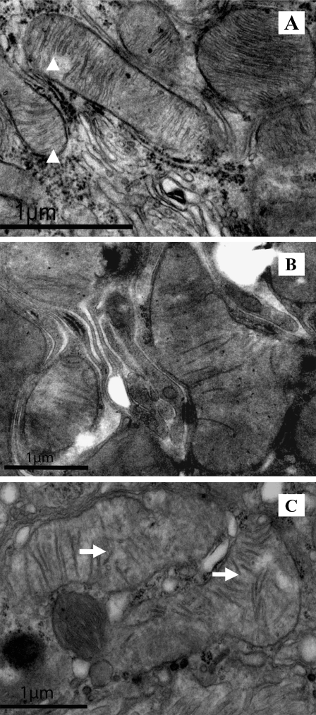
Autophagy is a highly regulated intracellular process for the degradation of cytoplasmic components, especially protein aggregates and damaged organelles. It is essential for maintaining healthy cells. Impaired or deficient autophagy is believed to cause or contribute to aging and age-related disease. In this study, we investigated the effects of age on autophagy in the kidneys of 3-, 12-, and 24-month-old Fischer 344 rats. The results revealed that autophagy-related gene (Atg)7 was significantly downregulated in kidneys of increasing age. The protein expression level of the autophagy marker light chain 3/Atg8 exhibited a marked decline in aged kidneys. The levels of p62/SQSTM1 and polyubiquitin aggregates, representing the function of autophagy and proteasomal degradation, increased in older kidneys. The level of 8-hydroxydeoxyguanosine, a marker of mitochondrial DNA oxidative damage, was also increased in older kidneys. Analysis by transmission electron microscope demonstrated swelling and disintegration of cristae in the mitochondria of aged kidneys. These results suggest that autophagic function decreases with age in the kidneys of Fischer 344 rats, and autophagy may mediate the process of kidney aging, leading to the accumulation of damaged mitochondria.
Figures

Similar articles
-
Impaired autophagic function in rat islets with aging.Age (Dordr). 2013 Oct;35(5):1531-44. doi: 10.1007/s11357-012-9456-0. Epub 2012 Jul 28. Age (Dordr). 2013. PMID: 22843415 Free PMC article.
-
Mitochondrial autophagy involving renal injury and aging is modulated by caloric intake in aged rat kidneys.PLoS One. 2013;8(7):e69720. doi: 10.1371/journal.pone.0069720. Epub 2013 Jul 22. PLoS One. 2013. PMID: 23894530 Free PMC article.
-
Short-term calorie restriction protects against renal senescence of aged rats by increasing autophagic activity and reducing oxidative damage.Mech Ageing Dev. 2013 Nov-Dec;134(11-12):570-9. doi: 10.1016/j.mad.2013.11.006. Epub 2013 Nov 27. Mech Ageing Dev. 2013. PMID: 24291536
-
Mitochondrial damage-induced impairment of angiogenesis in the aging rat kidney.Lab Invest. 2011 Feb;91(2):190-202. doi: 10.1038/labinvest.2010.175. Epub 2010 Oct 4. Lab Invest. 2011. PMID: 20921951
-
Autophagy, mitochondria and oxidative stress: cross-talk and redox signalling.Biochem J. 2012 Jan 15;441(2):523-40. doi: 10.1042/BJ20111451. Biochem J. 2012. PMID: 22187934 Free PMC article. Review.
Cited by
-
Mechanisms of Age-Dependent Loss of Dietary Restriction Protective Effects in Acute Kidney Injury.Cells. 2018 Oct 22;7(10):178. doi: 10.3390/cells7100178. Cells. 2018. PMID: 30360430 Free PMC article.
-
Treadmill Exercise Training Ameliorates Functional and Structural Age-Associated Kidney Changes in Male Albino Rats.ScientificWorldJournal. 2021 Nov 30;2021:1393372. doi: 10.1155/2021/1393372. eCollection 2021. ScientificWorldJournal. 2021. PMID: 34887703 Free PMC article.
-
Mitochondrial impairment in the five-sixth nephrectomy model of chronic renal failure: proteomic approach.BMC Nephrol. 2013 Oct 4;14:209. doi: 10.1186/1471-2369-14-209. BMC Nephrol. 2013. PMID: 24090408 Free PMC article.
-
Dietary Restriction for Kidney Protection: Decline in Nephroprotective Mechanisms During Aging.Front Physiol. 2021 Jul 6;12:699490. doi: 10.3389/fphys.2021.699490. eCollection 2021. Front Physiol. 2021. PMID: 34295266 Free PMC article. Review.
-
Targeting Premature Renal Aging: from Molecular Mechanisms of Cellular Senescence to Senolytic Trials.Front Pharmacol. 2021 Apr 29;12:630419. doi: 10.3389/fphar.2021.630419. eCollection 2021. Front Pharmacol. 2021. PMID: 33995028 Free PMC article. Review.
References
Publication types
MeSH terms
Substances
LinkOut - more resources
Full Text Sources
Medical