Mechanical hypersensitivity, sympathetic sprouting, and glial activation are attenuated by local injection of corticosteroid near the lumbar ganglion in a rat model of neuropathic pain
- PMID: 21455091
- PMCID: PMC3076946
- DOI: 10.1097/AAP.0b013e318203087f
Mechanical hypersensitivity, sympathetic sprouting, and glial activation are attenuated by local injection of corticosteroid near the lumbar ganglion in a rat model of neuropathic pain
Abstract
Background and objectives: Inflammatory responses in the lumbar dorsal root ganglion (DRG) play a key role in pathologic pain states. Systemic administration of a common anti-inflammatory corticosteroid, triamcinolone acetonide (TA), reduces sympathetic sprouting, mechanical pain behavior, spontaneous bursting activity, and cytokine and nerve growth factor production in the DRG. We hypothesized that systemic TA effects are primarily due to local effects on the DRG.
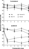
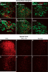
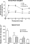
Methods: Male Sprague-Dawley rats were divided into 4 groups: SNL (tight ligation and transection of spinal nerves) and normal with and without a single dose of TA injectable suspension slowly injected onto the surface of DRG and surrounding region at the time of SNL or sham surgery. Mechanical threshold was tested on postoperative days 1, 3, 5, and 7. Immunohistochemical staining examined tyrosine hydroxylase and glial fibrillary acidic protein in DRG and CD11B antibody (OX-42) in spinal cord.
Results: Local TA treatment attenuated mechanical sensitivity, reduced sympathetic sprouting in the DRG, and decreased satellite glia activation in the DRG and microglia activation in the spinal cord after SNL.
Conclusions: A single injection of corticosteroid in the vicinity of the axotomized DRG can mimic many effects of systemic TA, mitigating behavioral and cellular abnormalities induced by spinal nerve ligation. This provides a further rationale for the use of localized steroid injections clinically and provides further support for the idea that localized inflammation at the level of the DRG is an important component of the spinal nerve ligation model, commonly classified as neuropathic pain model.
Figures


Similar articles
-
Early blockade of injured primary sensory afferents reduces glial cell activation in two rat neuropathic pain models.Neuroscience. 2009 Jun 2;160(4):847-57. doi: 10.1016/j.neuroscience.2009.03.016. Epub 2009 Mar 19. Neuroscience. 2009. PMID: 19303429 Free PMC article.
-
Systemic antiinflammatory corticosteroid reduces mechanical pain behavior, sympathetic sprouting, and elevation of proinflammatory cytokines in a rat model of neuropathic pain.Anesthesiology. 2007 Sep;107(3):469-77. doi: 10.1097/01.anes.0000278907.37774.8d. Anesthesiology. 2007. PMID: 17721250 Free PMC article.
-
Mirror-image pain is mediated by nerve growth factor produced from tumor necrosis factor alpha-activated satellite glia after peripheral nerve injury.Pain. 2014 May;155(5):906-920. doi: 10.1016/j.pain.2014.01.010. Epub 2014 Jan 18. Pain. 2014. PMID: 24447514
-
The dorsal root ganglion in chronic pain and as a target for neuromodulation: a review.Neuromodulation. 2015 Jan;18(1):24-32; discussion 32. doi: 10.1111/ner.12247. Epub 2014 Oct 29. Neuromodulation. 2015. PMID: 25354206 Review.
-
Modulating the delicate glial-neuronal interactions in neuropathic pain: promises and potential caveats.Neurosci Biobehav Rev. 2014 Sep;45:19-27. doi: 10.1016/j.neubiorev.2014.05.002. Epub 2014 May 10. Neurosci Biobehav Rev. 2014. PMID: 24820245 Free PMC article. Review.
Cited by
-
Effects of Long-Term Endogenous Corticosteroid Exposure on Brain Volume and Glial Cells in the AdKO Mouse.Front Neurosci. 2021 Feb 10;15:604103. doi: 10.3389/fnins.2021.604103. eCollection 2021. Front Neurosci. 2021. PMID: 33642975 Free PMC article.
-
Role of transforaminal epidural injections or selective nerve root blocks in the management of lumbar radicular syndrome - A narrative, evidence-based review.J Clin Orthop Trauma. 2020 Sep-Oct;11(5):802-809. doi: 10.1016/j.jcot.2020.06.004. Epub 2020 Jun 26. J Clin Orthop Trauma. 2020. PMID: 32904233 Free PMC article.
-
The Delayed-Onset Mechanical Pain Behavior Induced by Infant Peripheral Nerve Injury Is Accompanied by Sympathetic Sprouting in the Dorsal Root Ganglion.Biomed Res Int. 2020 Jun 16;2020:9165475. doi: 10.1155/2020/9165475. eCollection 2020. Biomed Res Int. 2020. PMID: 32626770 Free PMC article.
-
Increased Expression of Thymic Stromal Lymphopoietin in Chronic Constriction Injury of Rat Nerve.Int J Mol Sci. 2021 Jul 1;22(13):7105. doi: 10.3390/ijms22137105. Int J Mol Sci. 2021. PMID: 34281158 Free PMC article.
-
Acute morphine activates satellite glial cells and up-regulates IL-1β in dorsal root ganglia in mice via matrix metalloprotease-9.Mol Pain. 2012 Mar 22;8:18. doi: 10.1186/1744-8069-8-18. Mol Pain. 2012. PMID: 22439811 Free PMC article.
References
-
- Moalem G, Tracey DJ. Immune and inflammatory mechanisms in neuropathic pain. Brain Res Brain Res Rev. 2006;51:240–64. - PubMed
-
- DeLeo JA, Tanga FY, Tawfik VL. Neuroimmune activation and neuroinflammation in chronic pain and opioid tolerance/hyperalgesia. Neuroscientist. 2004;10:40–52. - PubMed
-
- Hanani M. Satellite glial cells in sensory ganglia: from form to function. Brain Res Brain Res Rev. 2005;48:457–76. - PubMed
Publication types
MeSH terms
Substances
Grants and funding
LinkOut - more resources
Full Text Sources
Other Literature Sources
Medical
Research Materials
Miscellaneous
