. 2011 May 4;30(9):1742-52.
doi: 10.1038/emboj.2011.85. Epub 2011 Mar 29.

T-cell receptor-induced JNK activation requires proteolytic inactivation of CYLD by MALT1

Affiliations

T-cell receptor-induced JNK activation requires proteolytic inactivation of CYLD by MALT1

Jens Staal et al. EMBO J. .

Abstract

The paracaspase mucosa-associated lymphoid tissue 1 (MALT1) is central to lymphocyte activation and lymphomagenesis. MALT1 mediates antigen receptor signalling to NF-κB by acting as a scaffold protein. Furthermore, MALT1 has proteolytic activity that contributes to optimal NF-κB activation by cleaving the NF-κB inhibitor A20. Whether MALT1 protease activity is involved in other signalling pathways, and the identity of the relevant substrates, is unknown. Here, we show that T-cell receptors (TCR) activation, as well as overexpression of the oncogenic API2-MALT1 fusion protein, results in proteolytic inactivation of CYLD by MALT1, which is specifically required for c-jun N-terminal kinase (JNK) activation and the inducible expression of a subset of genes. These results indicate a novel role for MALT1 proteolytic activity in TCR-induced JNK activation and reveal CYLD cleavage as the underlying mechanism.


Conflict of interest statement

The authors declare that they have no conflict of interest.

Figures
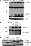
Figure 1
Figure 1
CYLD is cleaved upon T-cell and B-cell activation. (A) Jurkat cells were stimulated for the indicated times (minutes) with anti-CD3 plus anti-CD28. CYLD, IκBα (as a read-out for NF-κB activation), and actin (loading control) were analysed by immunoblotting. The CYLD antibody that was used for detection recognizes an N-terminal epitope and detects an N-terminal fragment of CYLD (CYLD-Nt). *Non-specific band. (B) Jurkat cells were stimulated overnight with anti-CD3 plus anti-CD28 or PMA plus ionomycin (PMA/I) and analysed for CYLD expression by immunoblotting (NS, non-stimulated cells). The CYLD antibody that was used for detection recognizes a C-terminal epitope and detects a C-terminal fragment of CYLD (CYLD-Ct). To illustrate the specificity of the signal, Jurkat cells were transfected with CYLD specific (CYLDi) and control (CTRi) siRNA 2 and 3 days prior to stimulation. *Non-specific band. (C) The B-cell lines SSK41 and Raji were stimulated for 1 h with anti-IgM or PMA plus ionomycin and analysed by immunoblotting with a CYLD antibody raised against C-terminal fragment of CYLD. Data are representative of three independent experiments.
Figure 2
Figure 2
CYLD cleavage requires MALT1 catalytic activity. (A) Effect of MALT1 silencing. (Top) Immunoblot analysis of CYLD cleavage in Jurkat cells transfected with MALT1-specific siRNA or control siRNA twice, 2 and 3 days prior to stimulation for 1 h with PMA plus ionomycin. CYLD was detected with an antibody raised against the C-terminus and recognizes the C-terminal fragment (CYLD-Ct). (Middle) Immunoblotting for MALT1 to demonstrate MALT1 silencing efficiency. (Bottom) Immunoblot analysis of β-actin (loading control). (B) Effect of an MALT1 peptide inhibitor. Immunoblot analysis of CYLD and A20 cleavage in Jurkat cells stimulated for 1 h with PMA plus ionomycine in the presence or absence of 100 μM of the MALT1 inhibitor z-VRPR-fmk, which was administered to the cells 1 h prior to stimulation. CYLD was detected with an antibody raised against the C-terminus or N-terminus. A20 was detected with an antibody raised against the C-terminus. (Bottom) Immunoblot analysis of β-actin (loading control). (C) Effect in primary T cells derived from MALT1-deficient mice. Primary CD4+ T cells derived from spleens of MALT1 wild-type (+/+) and MALT1-deficient (−/−) mice by MACS separation were stimulated with PMA plus ionomycin for 30 or 60 min and analysed with an antibody raised against the C-terminal fragment of CYLD (top). (Middle) MALT1 deficiency was confirmed by immunoblotting with anti-MALT1 (middle). (Bottom) Immunoblot analysis of β-actin (loading control). (D) Effect of overexpression of MALT1 in HEK293T cells. Cells were transiently transfected with N-terminally E-tagged CYLD and either wild-type MALT1 or catalytically inactive MALT1–C464A plus Bcl10 as indicated (lanes 1 and 2). Similarly, cells were transfected with API2–MALT1 or the catalytically inactive mutant API2–MALT1–C464A (lanes 3 and 4). CYLD cleavage was detected by immunoblotting with anti-E-tag. Data are representative of at least two independent experiments.
Figure 3
Figure 3
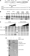
CYLD is directly cleaved by MALT1 at R324. (A) Cartoon of the domain structure of CYLD showing the three N-terminal CAP-Gly domains and the C-terminal DUB domain, as well as the amino-acid sequence of the region between the second and third CAP-Gly domains of human and mouse CYLD, respectively. The MALT1 cleavage site is indicated. (B) Mapping of the CYLD cleavage site. (Right) HEK293T cells were transfected with Flag-tagged human wild-type CYLD or the indicated human CYLD mutants together with API2–MALT1 (WT) or its inactive mutant API2–MALT1–C464A (C/A). Immunoblot analysis was performed with the CYLD-specific antibody raised against the C-terminus of CYLD. (C) In vitro cleavage of [S35]methionine-labelled CYLD (left) or CYLD–R324A (right) by increasing concentrations (0, 250, 500, and 1000 nM; wedges) of recombinant MALT1C–GyraseB, incubated for 1.5 h at 37°C, and analysed by SDS–PAGE and autoradiography. Data are representative of two independent experiments. (D) In vitro cleavage of recombinant CYLD by recombinant MALT1C–GyraseB, incubated for 1.5 h at 37°C, and analysed by SDS–PAGE and coomassie staining.
Figure 4
Figure 4
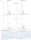
Determination of the CYLD cleavage site by LC–MS/MS. Recombinant CYLD was treated with recombinant MALT1C–GyraseB (5 μg) in the presence of 18O-labelled water (20% v/v of 93.7% H218O in reaction buffer), incubated for 1.5 h at 37°C, and analysed by SDS–PAGE. Gel-pieces of the full length and resulting proteolytic fragments were cut out and processed as described in the Materials and methods section. (A) MS spectra of representative peptide ions (doubly charged) contained in the N-terminal proteolytic CYLD fragment. The isotopic envelope distributions of the peptides derived from full-length CYLD or the N-terminal proteolytic CYLD fragment, here identified as 137-SGEEKFPGVVR-147, are indistinguishable, hinting that these peptides were not generated upon MALT1C–GyraseB-mediated CYLD proteolysis. (B) The peptide identified as 319-LAFMSR-324 is representative of the MALT1C–GyraseB generated neo-C-terminus in CYLD, as the difference in isotopic envelope distributions are indicative for the stable isotopic incorporation of 18O at the newly exposed carboxyl function of arginine 324 generated upon proteolysis. The extent of LAFMSR(18O), as determined by MS-isotope pattern calculator (http://prospector.ucsf.edu), was calculated 16%. (C) Obtained peptide coverage information of CYLD is highlighted in blue. The N-terminal His-tag is indicated in bold, the thrombin protease cleavage site (ENLYFQ▾G) underlined and the tryptic reporter-peptide indicative for MALT-1-specific cleavage at arginine 324 is in blue and underlined.
Figure 5
Figure 5
MALT1 proteolytic activity and CYLD cleavage are required for JNK signalling. (A) JNK and NF-κB signalling in CYLD silenced Jurkat cells. Immunoblot analysis of CYLD, IκBα, phospho-JNK (P-JNK), JNK, and β-actin from cells that are treated for the indicated times (minutes) with anti-CD3 plus anti-CD28. CYLD was silenced by transfection of CYLD-specific siRNA (CYLDi) or control siRNA (CTRi) 2 and 3 days prior to stimulation. (B) JNK and NF-κB signalling in Jurkat cells transfected with non-cleavable CYLD–R324A. Immunoblot analysis of CYLD, IκBα, phospho-JNK (P-JNK), JNK, and β-actin from anti-CD3 plus anti-CD28-stimulated cells that stably express wild-type CYLD or non-cleavable CYLD–R324A. The cleavage fragment in CYLD–R324A expressing cells results from endogenous CYLD expression. Identical results were obtained with several independent clones showing comparable expression of wild-type and CYLD–R324A. (C) p38 and ERK signalling in Jurkat cells transfected with non-cleavable CYLD–R324A. Immunoblot analysis of phospho-p38 (P-p38), p38, phospho-ERK (P-ERK), or β-actin from Jurkat cells that stably express wild-type CYLD or uncleavable CYLD–R324A. (D) JNK signalling in Jurkat cells in the presence of a MALT1 protease inhibitor. Immunoblot analysis of phospho-JNK (P-JNK), JNK, and β-actin from cells treated for the indicated times (minutes) with anti-CD3 plus anti-CD28. CYLD was silenced by transfection of CYLD-specific siRNA (CYLDi) or control siRNA (CTRi) 2 and 3 days prior to stimulation. Where indicated, cells received 100 μM of the MALT1 inhibitor z-VRPR-fmk 1 h prior to stimulation. Samples were loaded on a single gel. Data are representative of two (C, D) or three (A, B) independent experiments.
Figure 6
Figure 6
CYLD cleavage is required for TCR-induced IL-2 production in Jurkat cells. (A) IL-2 secretion. Jurkat cells stably expressing either wild-type CYLD or uncleavable CYLD–R324A were stimulated with anti-CD3 plus anti-CD28 (left) or PMA plus ionomycine (right) for 16 h, and the amount of IL-2 in the supernatant was analysed by ELISA. Endogenous CYLD was silenced with CYLD-specific morpholinos that were electroporated in the cells 2 and 3 days prior to stimulation. ELISA was performed in duplicate. Values and s.d. represent results from two independent duplicate experiments. (B) CYLD cleavage. Immunoblotting for CYLD of the cells used in (A). Data are representative of two independent experiments.
Figure 7
Figure 7
CYLD cleavage is required for TCR-induced expression of a subset of genes in Jurkat cells. (A) Jurkat cells stably expressing either wild-type CYLD (black line with ▪ symbol) or uncleavable CYLD–R324A (grey line with x symbol) were stimulated with PMA plus ionomycine (A) or anti-CD3 plus anti-CD28 (B) for the times indicated (minutes), and IL-2, IL-8, A20, IκBα, and c-Jun expression were determined by Q-PCR and normalized against the two house-keeping genes HPRT1 and PPIA. Data are representative of three independent experiments.
Figure 8
Figure 8
Scheme illustrating our model for the role of MALT1-mediated CYLD cleavage in TCR-induced JNK activation. TCR stimulation or direct activation of PKC by the phorbol ester TPA results in the activation of the CARMA1/BCL10/MALT1 complex, leading to IKK- and JNK-mediated activation of NF-κB and AP-1 transcription factors, respectively. CYLD is negatively regulating both signalling pathways. (Left panel) MALT1 rapidly cleaves CYLD (as part of a signalling complex that specifically controls JNK activation), preventing it from blocking JNK activation. (Right panel) MALT1 cannot cleave the CYLD–R324A mutant, which can now fully inhibit JNK activation.

Similar articles

Cited by

References

    1. Angel P, Hattori K, Smeal T, Karin M (1988) The jun proto-oncogene is positively autoregulated by its product, Jun/AP-1. Cell 55: 875–885 - PubMed
    1. Arnesen T, Starheim K, Van Damme P, Evjenth R, Betts M, Ryningen A, Vandekerckhove J, Gevaert K, Anderson D (2010) The chaperone-like protein HYPK acts together with NatA in cotranslational N-terminal acetylation and prevention of Huntingtin aggregation. Mol Cell Biol 30: 1898–1909 - PMC - PubMed
    1. Blonska M, Pappu BP, Matsumoto R, Li H, Su B, Wang D, Lin X (2007) The CARMA1-Bcl10 signaling complex selectively regulates JNK2 kinase in the T cell receptor-signaling pathway. Immunity 26: 55–66 - PMC - PubMed
    1. Coornaert B, Baens M, Heyninck K, Bekaert T, Haegman M, Staal J, Sun L, Chen ZJ, Marynen P, Beyaert R (2008) T cell antigen receptor stimulation induces MALT1 paracaspase-mediated cleavage of the NF-κB inhibitor A20. Nat Immunol 9: 263–271 - PubMed
    1. Coornaert B, Carpentier I, Beyaert R (2009) A20: central gatekeeper in inflammation and immunity. J Biol Chem 284: 8217–8221 - PMC - PubMed

Publication types

MeSH terms