The development and potential of acoustic radiation force impulse (ARFI) imaging for carotid artery plaque characterization
- PMID: 21447606
- PMCID: PMC3265036
- DOI: 10.1177/1358863X11400936
The development and potential of acoustic radiation force impulse (ARFI) imaging for carotid artery plaque characterization
Abstract
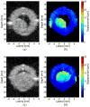
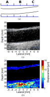
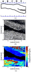
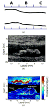
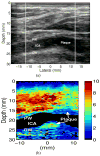
Stroke is the third leading cause of death and long-term disability in the USA. Currently, surgical intervention decisions in asymptomatic patients are based upon the degree of carotid artery stenosis. While there is a clear benefit of endarterectomy for patients with severe (> 70%) stenosis, in those with high/moderate (50-69%) stenosis the evidence is less clear. Evidence suggests ischemic stroke is associated less with calcified and fibrous plaques than with those containing softer tissue, especially when accompanied by a thin fibrous cap. A reliable mechanism for the identification of individuals with atherosclerotic plaques which confer the highest risk for stroke is fundamental to the selection of patients for vascular interventions. Acoustic radiation force impulse (ARFI) imaging is a new ultrasonic-based imaging method that characterizes the mechanical properties of tissue by measuring displacement resulting from the application of acoustic radiation force. These displacements provide information about the local stiffness of tissue and can differentiate between soft and hard areas. Because arterial walls, soft tissue, atheromas, and calcifications have a wide range in their stiffness properties, they represent excellent candidates for ARFI imaging. We present information from early phantom experiments and excised human limb studies to in vivo carotid artery scans and provide evidence for the ability of ARFI to provide high-quality images which highlight mechanical differences in tissue stiffness not readily apparent in matched B-mode images. This allows ARFI to identify soft from hard plaques and differentiate characteristics associated with plaque vulnerability or stability.
Figures




Similar articles
-
Non-invasive in vivo characterization of human carotid plaques with acoustic radiation force impulse ultrasound: comparison with histology after endarterectomy.Ultrasound Med Biol. 2015 Mar;41(3):685-97. doi: 10.1016/j.ultrasmedbio.2014.09.016. Epub 2015 Jan 22. Ultrasound Med Biol. 2015. PMID: 25619778 Free PMC article.
-
Comparison of Acoustic Radiation Force Impulse Imaging Derived Carotid Plaque Stiffness With Spatially Registered MRI Determined Composition.IEEE Trans Med Imaging. 2015 Nov;34(11):2354-65. doi: 10.1109/TMI.2015.2432797. Epub 2015 May 13. IEEE Trans Med Imaging. 2015. PMID: 25974933 Free PMC article.
-
Acoustic radiation force impulse imaging for noninvasive characterization of carotid artery atherosclerotic plaques: a feasibility study.Ultrasound Med Biol. 2009 May;35(5):707-16. doi: 10.1016/j.ultrasmedbio.2008.11.001. Epub 2009 Feb 25. Ultrasound Med Biol. 2009. PMID: 19243877 Free PMC article.
-
Review: Mechanical Characterization of Carotid Arteries and Atherosclerotic Plaques.IEEE Trans Ultrason Ferroelectr Freq Control. 2016 Oct;63(10):1613-1623. doi: 10.1109/TUFFC.2016.2572260. Epub 2016 May 26. IEEE Trans Ultrason Ferroelectr Freq Control. 2016. PMID: 27249826 Review.
-
Contemporary carotid imaging: from degree of stenosis to plaque vulnerability.J Neurosurg. 2016 Jan;124(1):27-42. doi: 10.3171/2015.1.JNS142452. Epub 2015 Jul 31. J Neurosurg. 2016. PMID: 26230478 Review.
Cited by
-
Intravascular polarization-sensitive optical coherence tomography based on polarization mode delay.Sci Rep. 2022 Apr 27;12(1):6831. doi: 10.1038/s41598-022-10709-8. Sci Rep. 2022. PMID: 35477738 Free PMC article.
-
A direct vulnerable atherosclerotic plaque elasticity reconstruction method based on an original material-finite element formulation: theoretical framework.Phys Med Biol. 2013 Dec 7;58(23):8457-76. doi: 10.1088/0031-9155/58/23/8457. Epub 2013 Nov 15. Phys Med Biol. 2013. PMID: 24240392 Free PMC article.
-
Viscoelastic response (VisR) imaging for assessment of viscoelasticity in Voigt materials.IEEE Trans Ultrason Ferroelectr Freq Control. 2013 Dec;60(12):2488-500. doi: 10.1109/TUFFC.2013.2848. IEEE Trans Ultrason Ferroelectr Freq Control. 2013. PMID: 24297015 Free PMC article.
-
Characterisation of carotid plaques with ultrasound elastography: feasibility and correlation with high-resolution magnetic resonance imaging.Eur Radiol. 2013 Jul;23(7):2030-41. doi: 10.1007/s00330-013-2772-7. Epub 2013 Feb 17. Eur Radiol. 2013. PMID: 23417249
-
Methods for robust in vivo strain estimation in the carotid artery.Phys Med Biol. 2012 Nov 21;57(22):7329-53. doi: 10.1088/0031-9155/57/22/7329. Epub 2012 Oct 18. Phys Med Biol. 2012. PMID: 23079725 Free PMC article.
References
-
- Rosamond W, Flegal K, Friday G, Furie K, Go A, Greenlund K, Haase N, Ho M, Howard V, Kissela B, Kittner S, Lloyd-Jones D, McDermott M, Meigs J, Moy C, Nichol G, O’Donnell CJ, Roger V, Rumsfeld J, Sorlie P, Steinberger J, Thom T, Wasserthiel-Smoller S, Hong Y for the American Heart Association Statistics C, Stroke Statistics S. Heart disease and stroke statistics--2007 update: A report from the american heart association statistics committee and stroke statistics subcommittee. Circulation. 2007;115:e69–171. - PubMed
-
- Timsit S, Sacco R, Mohr J, Foulkes M, Tatemichi T, Wolf P, Price T, Hier D. Early clinical differentiation of cerebral infarction from severe atherosclerotic stenosis and cardioembolism. Stroke. 2007;23:486–491. - PubMed
-
- North American Symptomatic Carotid Endarterectomy Trial C. Beneficial effect of carotid endarterectomy in symptomatic patients with high-grade carotid stenosis. N Engl J Med. 1991;325:445–453. - PubMed
-
- Warlow C. Mrc european carotid surgery trial: Interim results for symptomatic patients with severe (70–99%) or with mild (0–29%) carotid stenosis. The Lancet. 1991;337:1235–1243. - PubMed
-
- Study ECftACA. Endarterectomy for asymptomatic carotid artery stenosis. JAMA. 1995;273:1421–1428. - PubMed
Publication types
MeSH terms
Grants and funding
LinkOut - more resources
Full Text Sources
Other Literature Sources
Miscellaneous

