Regulation of AKT phosphorylation at Ser473 and Thr308 by endoplasmic reticulum stress modulates substrate specificity in a severity dependent manner
- PMID: 21445305
- PMCID: PMC3061875
- DOI: 10.1371/journal.pone.0017894
Regulation of AKT phosphorylation at Ser473 and Thr308 by endoplasmic reticulum stress modulates substrate specificity in a severity dependent manner
Abstract
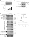
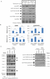
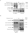
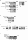
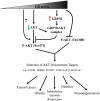
Endoplasmic reticulum (ER) stress is a common factor in the pathophysiology of diverse human diseases that are characterised by contrasting cellular behaviours, from proliferation in cancer to apoptosis in neurodegenerative disorders. Coincidently, dysregulation of AKT/PKB activity, which is the central regulator of cell growth, proliferation and survival, is often associated with the same diseases. Here, we demonstrate that ER stress modulates AKT substrate specificity in a severity-dependent manner, as shown by phospho-specific antibodies against known AKT targets. ER stress also reduces both total and phosphorylated AKT in a severity-dependent manner, without affecting activity of the upstream kinase PDK1. Normalisation to total AKT revealed that under ER stress phosphorylation of Thr308 is suppressed while that of Ser473 is increased. ER stress induces GRP78, and siRNA-mediated knock-down of GRP78 enhances phosphorylation at Ser473 by 3.6 fold, but not at Thr308. Substrate specificity is again altered. An in-situ proximity ligation assay revealed a physical interaction between GRP78 and AKT at the plasma membrane of cells following induction of ER stress. Staining was weak in cells with normal nuclear morphology but stronger in those displaying rounded, condensed nuclei. Co-immunoprecipitation of GRP78 and P-AKT(Ser473) confirmed the immuno-complex consists of non-phosphorylated AKT (Ser473 and Thr308). The interaction is likely specific as AKT did not bind to all molecular chaperones, and GRP78 did not bind to p70 S6 kinase. These findings provide one mechanistic explanation for how ER stress contributes to human pathologies demonstrating contrasting cell fates via modulation of AKT signalling.
Conflict of interest statement
Figures

Similar articles
-
AKT phosphorylation sites of Ser473 and Thr308 regulate AKT degradation.Biosci Biotechnol Biochem. 2019 Mar;83(3):429-435. doi: 10.1080/09168451.2018.1549974. Epub 2018 Nov 29. Biosci Biotechnol Biochem. 2019. PMID: 30488766
-
Tamoxifen-induced cytotoxicity in breast cancer cells is mediated by glucose-regulated protein 78 (GRP78) via AKT (Thr308) regulation.Int J Biochem Cell Biol. 2016 Aug;77(Pt A):57-67. doi: 10.1016/j.biocel.2016.05.021. Epub 2016 Jun 1. Int J Biochem Cell Biol. 2016. PMID: 27262235
-
Dephosphorylation and inactivation of Akt/PKB is counteracted by protein kinase CK2 in HEK 293T cells.Cell Mol Life Sci. 2009 Oct;66(20):3363-73. doi: 10.1007/s00018-009-0108-1. Epub 2009 Aug 8. Cell Mol Life Sci. 2009. PMID: 19662498 Free PMC article.
-
The potential role of Akt phosphorylation in human cancers.Int J Biol Markers. 2008 Jan-Mar;23(1):1-9. doi: 10.1177/172460080802300101. Int J Biol Markers. 2008. PMID: 18409144 Review.
-
Control of Akt activity and substrate phosphorylation in cells.IUBMB Life. 2020 Jun;72(6):1115-1125. doi: 10.1002/iub.2264. Epub 2020 Mar 3. IUBMB Life. 2020. PMID: 32125765 Free PMC article. Review.
Cited by
-
The Interplay Between Autophagy and Regulated Necrosis.Antioxid Redox Signal. 2023 Mar;38(7-9):550-580. doi: 10.1089/ars.2022.0110. Epub 2022 Oct 12. Antioxid Redox Signal. 2023. PMID: 36053716 Free PMC article. Review.
-
Bromodomain inhibitor jq1 induces cell cycle arrest and apoptosis of glioma stem cells through the VEGF/PI3K/AKT signaling pathway.Int J Oncol. 2019 Oct;55(4):879-895. doi: 10.3892/ijo.2019.4863. Epub 2019 Aug 29. Int J Oncol. 2019. PMID: 31485609 Free PMC article.
-
New Insights into Protein Kinase B/Akt Signaling: Role of Localized Akt Activation and Compartment-Specific Target Proteins for the Cellular Radiation Response.Cancers (Basel). 2018 Mar 18;10(3):78. doi: 10.3390/cancers10030078. Cancers (Basel). 2018. PMID: 29562639 Free PMC article. Review.
-
Remote Ischemic Preconditioning induces Cardioprotective Autophagy and Signals through the IL-6-Dependent JAK-STAT Pathway.Int J Mol Sci. 2020 Mar 1;21(5):1692. doi: 10.3390/ijms21051692. Int J Mol Sci. 2020. PMID: 32121587 Free PMC article.
-
Modulation of endothelial cell migration by ER stress and insulin resistance: a role during maternal obesity?Front Pharmacol. 2014 Aug 19;5:189. doi: 10.3389/fphar.2014.00189. eCollection 2014. Front Pharmacol. 2014. PMID: 25191269 Free PMC article. Review.
References
-
- Rutkowski DT, Kaufman RJ. A trip to the ER: coping with stress. Trends Cell Biol. 2004;14:20–28. - PubMed
-
- Kim R, Emi M, Tanabe K, Murakami S. Role of the unfolded protein response in cell death. Apoptosis. 2006;11:5–13. - PubMed
-
- Gonzalez-Gronow M, Selim MA, Papalas J, Pizzo SV. GRP78: a multifunctional receptor on the cell surface. Antioxid Redox Signal. 2009;11:2299–2306. - PubMed
Publication types
MeSH terms
Substances
Grants and funding
LinkOut - more resources
Full Text Sources
Molecular Biology Databases
Miscellaneous

