. 2011 Jun;133(2):206-20.
doi: 10.1111/j.1365-2567.2011.03428.x. Epub 2011 Mar 23.

Development of an attenuated interleukin-2 fusion protein that can be activated by tumour-expressed proteases

Affiliations

Development of an attenuated interleukin-2 fusion protein that can be activated by tumour-expressed proteases

John Puskas et al. Immunology. 2011 Jun.

Abstract

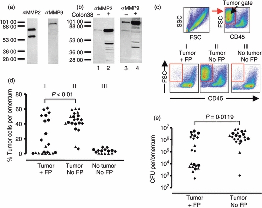
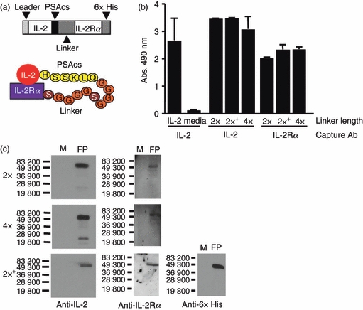
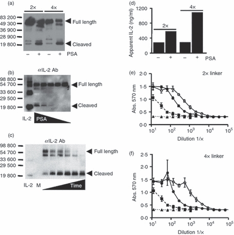
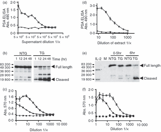
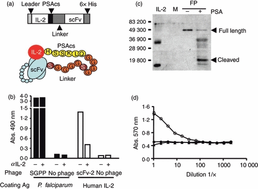
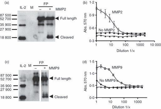
The ability to alter the cytokine microenvironment has the potential to shape immune responses in many physiological settings, including the immunotherapy of tumours. We set out to develop a general approach in which cytokines could be functionally attenuated until activated. We report the development and initial characterization of fusion proteins in which human or mouse interleukin-2 (IL-2), a potent growth factor for immune cells, is joined to a specific IL-2 inhibitory binding component separated by a protease site. The rationale is that upon cleavage by a protease the cytokine is free to dissociate from the inhibitory component and becomes biologically more available. We describe the successful development of two attenuation strategies using specific binding: the first uses the mouse IL-2 receptor alpha chain as the inhibitory binding component whereas the second employs a human antibody fragment (scFv) reactive with human IL-2. We demonstrated that the fusion proteins containing a prostate-specific antigen or a matrix metalloproteinase (MMP) protease cleavage site are markedly attenuated in the intact fusion protein but had enhanced bioactivity of IL-2 in vitro when cleaved. Further, we showed that a fusion protein composed of the IL-2/IL-2 receptor alpha chain with an MMP cleavage site reduced tumour growth in vivo in a peritoneal mouse tumour model. This general strategy should be applicable to other proteases and immune modulators allowing site-specific activation of immunomodulators while reducing unwanted side-effects.


Figures

Figure 6
Figure 6
Fusion protein treatment reduces Colon 38 tumour growth in vivo. (a) In vitro expression of matrix metalloproteinase 2 (MMP2) and MMP9 using the Colon 38 tumour cell line. Immunoblot analyses of in vitro grown Colon 38 supernatants using the indicated antibodies. (b) In vivo expression of MMP2 and MMP9 from omental lysates with and without Colon 38 tumour present. Lanes 1 and 2 were probed with the anti-MMP2 antibody. Lane 1 contains omental lysate from an untreated mouse with no tumour. Lane 2 contains lysate from an omentum that had Colon 38 grown in vivo. Lanes 3 and 4 are replicates of lanes 1 and 2 except they were probed with an anti-MMP9 antibody. Dashes and numbers indicate apparent molecular weights. Note lower molecular weight bands probably represent cleavage products. (c) Gating scheme and representative flow analyses used to identify tumour cells (high forward scatter, CD45 negative) growing in vivo on the omentum. Reconstitution experiments in which tumour cells were added to omental cells were used to establish the tumour gate, which is indicated by the box. Bottom panels are examples of flow analyses of omenta from mice which received tumour and MMP fusion protein treatment (panel I: tumour +FP), mice which had received tumour but no treatment with MMP fusion protein (panel II: tumour No FP), or mice which received neither tumour nor fusion protein (panel III: no tumour, No FP). (d) Compiled analyses of tumour cells detected on the omentum by flow cytometry. Each symbol represents an individual mouse. Different symbols indicate mice from three experiments. The P-value between the indicated groups was calculated using the Kruskal–Wallis test. (e) Viable tumour cells were determined by colony-forming assay. Symbols indicate individual mice. P-value was calculated using the Mann–Whitney U-test.
Figure 1
Figure 1
Characterization of mouse IL-2/PSAcs/IL-2Rα fusion proteins. (a) Schematic diagram of mouse IL-2/PSAcs/linker/IL-2Rα fusion proteins. The prostate-specific antigen cleavage sequence (PSAcs) indicates the amino acid sequence HSSKLQ that can be cleaved by the PSA protease followed by the 2 × (GGGGS)2 (shown) or 4 × (GGGGS)4 linker lengths. (b) ELISA analysis of fusion proteins using capture antibodies to interleukin-2 (IL-2; JES6-1A12) or IL-2 receptor alpha chain (IL-2Rα; PC61) and a different biotin-labelled anti-IL-2-detecting antibody (JES5H4) showing that both IL-2 and the IL-2Rα moieties are present on the same molecule. 2 × indicates (GGGGS)2 linker, 4 × indicates (GGGGS)4 linker, and (+) indicates that the construct contains the 6 × His tag. (c) Immunoblot analyses of the fusion proteins using antibodies reactive with IL-2 (JES6-1A12), IL-2Rα (PC61) and 6 × His epitope tag (MM5-156P). Molecular weight standards are indicated. The full length fusion proteins (FP) have an approximate molecular weight of 50 000 MW and medium was run as a negative control (M).
Figure 2
Figure 2
Prostate-specific antigen (PSA) treatment enhances antibody accessibility and interleukin-2 (IL-2) functional activity in mouse IL-2/PSAcs/IL-2Rα fusion proteins. Fusion proteins (2 × and 4 × linker lengths) were treated with PSA or buffer controls for 1 hr at 37° and aliquots were analysed by immunoblot, ELISA, or by functional analysis. (a) Anti-IL-2 immunoblot analysis of fusion proteins before and after treatment with PSA. Bars indicate molecular weight markers, (+) indicates treatment with PSA, (−) indicates treatment with control buffer, and full length and predicted cleavage product containing IL-2 have been denoted (arrowheads). (b) Titration of PSA using mouse IL-2/PSAcs/2 × linker/IL-2Rα fusion protein at 37° for 1 hr and immunoblot analysis using an anti-mouse IL-2 antibody (JES6-1A12). Bars and numbers indicate molecular weight markers. Full length and cleaved fusion proteins have been denoted. The amount of PSA used in the reaction is as follows: 11·25, 5·6, 2·8, 1·4 and 0 μg. (c) PSA time–course using mouse IL-2/PSAcs/4 × linker/IL-2Rα fusion protein digested with 5 μg for 0, 1, 3, 6, 12 and 24 hr at 37° analysed by immunoblot with an anti-mouse IL-2 antibody (JES6-1A12). Medium was run as negative control (M). (d) An ELISA used to measure the amount of IL-2 before and after treatment with PSA. An apparent increase in IL-2 can be seen after PSA incubation for both fusion proteins tested. (e, f) Functional analyses of IL-2 before and after cleavage. Biologically active IL-2 from the fusion proteins was measured using the CTLL-2 functional assay. Fusion protein treated with PSA (○), or with buffer control (•), IL-2 standard (▪), and medium control (▴). The same amount of fusion protein was present in the PSA-treated and untreated samples used in the CTLL-2 assay. Points represent the average of three replicates and error bars indicate standard deviation. Representative of three independent experiments.
Figure 3
Figure 3
Interleukin-2 (IL-2) bioactivity increases when IL-2/PSAcs/4 × linker/IL-2Rα fusion protein is cultured with explanted prostates or homogenized prostate extracts. Prostates were removed from non-transgenic (NTG) or prostate-specific antigen (PSA) transgenic (TG) C57BL/6J mice, cultured at 37° with fusion protein and aliquots of media containing fusion protein were removed at 1, 12, 24, 48 hr for analysis. (a) Detection of PSA in TG (□) or NTG (▪) supernatant aliquots at 48 hr by ELISA. (b) Immunoblot analysis using an anti-IL-2 antibody of the culture supernatants at the indicated time-points. Bars and numbers indicate molecular weight markers. The full length and the predicted cleavage products containing IL-2 are indicated by arrowheads. (c) IL-2 functional assay at 48 hr. Supernatant from cultures containing TG prostate explants (○), NTG explants (•), media control (▪). The same amount of culture supernatant was used for the CTLL-2 assay. (d) Analysis of PSA by ELISA in homogenized prostate extracts from TG (○) and NTG (•) mice. The first well represents approximately 40 ng of NTG or TG extract. (b) Immunoblot analysis using an anti-IL-2 antibody of the samples containing prostate extracts and the fusion protein at 0·5 or 6 hr at 37°. Bacterially derived non-glycosylated recombinant mouse IL-2 (10 ng) was used as a standard and has an approximate molecular weight of 18 500 MW. The band at approximately 37 000 MW in the control lane probably represents a dimer of IL-2. Medium-only control (M). Full-length fusion protein and the predicted cleavage product containing IL-2 have been denoted by arrowheads. (f) IL-2 functional assay of fusion proteins after incubation for 6 hr at 37° with prostate extracts. TG prostate extracts (○), NTG extracts (•) and media control (▪). Equivalent amounts of fusion protein were present in the PSA-treated and untreated samples used in the CTLL-2 assay.
Figure 4
Figure 4
Characterization and analyses of human IL-2/PSAcs/scFv fusion proteins. (a) Schematic diagram of human IL-2/PSAcs/scFv fusion proteins containing human interleukin-2 (IL-2) fused to the prostate-specific antigen cleavage sequence (PSAcs), a (GGGGS)2 or (GGGGS)4 linker unit followed by VL and VH fragments of an antibody tethered together by a linker (scFv) and a 6 × His carboxyl tag. (b) A modified ELISA using scFv phage was performed and a phage clone expressing scFv (phscFv) that binds human IL-2 (scFv-2) was screened for the ability to be inhibited by the anti-human IL-2 neutralizing antibody (MQ1-17H12). Black columns indicate recombinant Plasmodium falciparum protein (SGPP) coating antigen, white columns indicate human IL-2 as the coating antigen. A phage which bound SGPP served as a control and this binding was not inhibited by the anti-human IL-2 neutralizing antibody whereas the phage clone scFv-2 could be partially blocked by the antibody. (c) Anti-human IL-2 immunoblot analysis of fusion protein treated with purified PSA or with control PSA buffer treatment. Bars and numbers indicate molecular weight markers. Full-length fusion protein (FP) and the predicted cleavage product containing IL-2 have been denoted with arrowheads. Media negative control (M). Bacterially derived non-glycosylated recombinant human IL-2 (50 ng) was used as a standard and has an approximate molecular weight of 15 500. The fusion protein IL-2 was derived from insect cells and may be post-translationally modified accounting for its slightly higher molecular weight. (d) IL-2 functional assay on fusion protein. Treatment with PSA (○), control buffer (•) or media control (▴). Equal amounts of fusion protein were used in the CTLL-2 assay.
Figure 5
Figure 5
Evaluation of mouse IL-2/MMPcs/4 × linker/IL-2Rα + 6 × His fusion proteins digested with matrix metalloproteinase 2 (MMP2) or MMP9. The fusion protein containing the MMP cleavage sequence was incubated with either MMP9 or MMP2 or buffer treated and the resulting material was tested by Western blotting analysis and for activity using the CTLL-2 assay. (a) Immunoblot analyses of the fusion protein digested with MMP2 using an anti-interleukin-2 (IL-2) antibody. Bars and numbers indicate molecular weight markers. The full length and the predicted cleavage product containing IL-2 are indicated by arrowheads. Media control (M). (b) Fusion protein digests were run in the CTLL-2 assay using equal amounts of fusion protein for the MMP2 treated or untreated fusion protein. Fusion protein plus MMP2 (○), fusion protein no treatment (•), media control (▴). (c) Immunoblot analyses of the fusion protein digested with MMP9 using anti-IL-2 antibody. Bars and numbers indicate molecular weight markers. The full length and the predicted cleavage product containing IL-2 are indicated by arrowheads. Note that the + and – MMP9 digests were run in the same gel and exposed for the same amount of time but some intervening lanes were removed. (d) Fusion protein digests were run in the CTLL-2 assay using equal amounts of fusion protein for the MMP9 treated or untreated fusion protein. Fusion protein plus MMP9 (○), fusion protein no teatment (•), media control (▴). Note: in some cases error bars are not visible in CTLL-2 assay because of the size of symbols.

Similar articles

Cited by

References

    1. Finn OJ. Cancer immunology. N Engl J Med. 2008;358:2704–15. - PubMed
    1. Boon T, Coulie PG, Van den Eynde BJ, van der Bruggen P. Human T cell responses against melanoma. Annu Rev Immunol. 2006;24:175–208. - PubMed
    1. Parmiani G, De Filippo A, Novellino L, Castelli C. Unique human tumor antigens: immunobiology and use in clinical trials. J Immunol. 2007;178:1975–9. - PubMed
    1. Berzofsky JA, Terabe M, Oh S, Belyakov IM, Ahlers JD, Janik JE, Morris JC. Progress on new vaccine strategies for the immunotherapy and prevention of cancer. J Clin Invest. 2004;113:1515–25. - PMC - PubMed
    1. Slingluff CL, Jr, Chianese-Bullock KA, Bullock TN, et al. Immunity to melanoma antigens: from self-tolerance to immunotherapy. Adv Immunol. 2006;90:243–95. - PubMed

Publication types

MeSH terms

LinkOut - more resources