. 2010 Sep 8:2:136.
doi: 10.3389/fnsyn.2010.00136. eCollection 2010.

Novel roles for immune molecules in neural development: implications for neurodevelopmental disorders

Affiliations

Novel roles for immune molecules in neural development: implications for neurodevelopmental disorders

Paula A Garay et al. Front Synaptic Neurosci. .

Abstract

Although the brain has classically been considered "immune-privileged", current research suggests an extensive communication between the immune and nervous systems in both health and disease. Recent studies demonstrate that immune molecules are present at the right place and time to modulate the development and function of the healthy and diseased central nervous system (CNS). Indeed, immune molecules play integral roles in the CNS throughout neural development, including affecting neurogenesis, neuronal migration, axon guidance, synapse formation, activity-dependent refinement of circuits, and synaptic plasticity. Moreover, the roles of individual immune molecules in the nervous system may change over development. This review focuses on the effects of immune molecules on neuronal connections in the mammalian central nervous system - specifically the roles for MHCI and its receptors, complement, and cytokines on the function, refinement, and plasticity of geniculate, cortical and hippocampal synapses, and their relationship to neurodevelopmental disorders. These functions for immune molecules during neural development suggest that they could also mediate pathological responses to chronic elevations of cytokines in neurodevelopmental disorders, including autism spectrum disorders (ASD) and schizophrenia.

Keywords: autism; complement; cytokine; major histocompatibility complex; plasticity; refinement; schizophrenia; synapse.


Figures
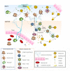
Figure 1
Figure 1
Innate and adaptive immunity. A simplified schematic of the two branches of the immune response. Following injury or infection, pathogens (bacteria, virus or foreign protein) infiltrate tissue. The innate response provides immediate defense against infection (1–5): 1. Neutrophils engulf the pathogen and destroy it by releasing antimicrobial toxins. 2. Macrophages can directly phagocytose pathogens, leading to production of cytokines and recruitment of more cells from the blood. 3. Infected cells displaying low levels of MHCI on their surface are directly detected by natural killer (NK) cells, which release lytic enzymes causing the infected cell to die via apoptosis. 4. Bacteria can also be recognized by the complement system, resulting in their lysis. 5. Macrophages and dendritic cells can become antigen presenting cells (APCs) by taking up peripheral antigens and migrating to lymph nodes to present antigen on their surface to naïve B- and T- cells. The adaptive response confers the ability to recognize and remember specific pathogens to generate immunity (6–11): 6. APC interaction with B- and T- cells in the lymph nodes leads to B- and T-cell activation and migration to the periphery where they mediate adaptive immunity. 7. Once activated, the T-cell undergoes a process of clonal expansion in which it divides rapidly to produce multiple identical effector cells. Activated T-cells then travel to the periphery in search of infected cells displaying cognate antigen/MHCI complex. 8. Peripheral APCs induce helper T cells to release cytokines and recruit cytotoxic T cells (CTL). 9. Activated antigen-specific B cells receiving signals from helper T-cells differentiate into plasma cells and secrete antibodies. 10. Antibodies bind to target antigens forming immune complexes which can then activate complement or be taken up by macrophages through Fc receptors 11. Formation of cytotoxic T-cell synapses causes lysis of the infected cell.
Figure 2
Figure 2
Immune and neuronal synapses. Highly simplified schematics of these synapses illustrate the common core components of these asymmetric junctions. (A) A cytotoxic T-cell immune synapse is formed between an antigen-specific T-cell and a cell infected with an intracellular pathogen. The close junction is formed from a ring of adhesion proteins (purple cylinders) surrounding an inner signaling molecular domain of antigen receptors (MHCI; gray y) bound to T-cell receptors (TCR; blue Y). Cytokine receptors also cluster in the synapse (not shown in the diagram) where they are exposed to cytokines secreted into the synapse. (B, left) Activation of TCR signaling by MHCI molecules presenting non-self antigens (orange ovals) causes a signaling cascade resulting in polarization of the actin and microtubule cytoskeleton (blue lines) in the T-cell, recruitment of lytic granules (circles) and cell-surface receptors and co-stimulatory molecules (including trans-synaptic adhesion molecules [purple ovals]) to the synapse, secretion of lytic granules (red) and apoptosis of the infected cell. (B, right) Conversely, TCR interaction with an MHC1 molecule containing a non-TCR specific peptide (orange ovals) does not affect microtubule reorganization and does not recruit lytic granules, resulting in termination of the contact and survival the APC. (C)The lytic natural killer (NK) cell immune synapse is also specialized for mediating cytotoxicity. An encounter between an NK cell and a target cell results in adhesion (orange ovals). The balance between activating and inhibitory receptor (pink) signaling at the cell–cell contact determines the outcome of the interaction. (D, left) A lack of MHCI on the target cell, caused by viral infection or tumorigenesis, triggers a signaling cascade in the NK cell resulting in reorganization of the actin cytoskeleton, clustering of cell-surface receptors (pink) and signaling molecules (blue) in the NK cell, recruitment and secretion of lytic granules, ultimately resulting in lysis of the target cell. (D, right) Conversely, the presence of MHCI (gray y) on the target cell results in binding of MHCI to NK inhibitory receptors, including PirB and Ly49s and initiates dominant inhibitory signaling, preventing the formation of the NK cell activation synapse and resulting in survival of the target cell. (E) Glutamatergic synapses in the mammalian CNS are comprised of several major protein classes. In the presynaptic axon terminal (blue), synaptic vesicles (circles) containing the neurotransmitter glutamate (blue) cycle at the active zone, which is composed of many kinds of proteins including presynaptic scaffolding proteins (blue Xs and hooks). The presynaptic terminal is separated from the postsynaptic dendrite (pink) by the synaptic cleft (yellow). A number of families of trans-synaptic adhesion molecules (blue ovals) span this cleft, providing a molecular connection capable of rapid signaling between the pre- and postsynaptic membranes. Glutamate receptors (gray), including AMPA and NMDA receptors, are found in the postsynaptic membrane, where they are associated with a large number of scaffolding and signaling proteins (pink rectangles) that together comprise the postsynaptic density. (F) If the synapse is weakened by long-term depression (LTD) then it can be eliminated (left), but if it is strengthened by long-term potentiation (LTP) then it will be stabilized and grow (right).
Figure 3
Figure 3
The relationship between the immune system and nervous system is highly complicated. However, it is now apparent that immune molecules not only cross the blood–brain barrier in times of injury, but are expressed during normal brain development. Recent evidence suggests roles for MHCI and its receptors, complement, and cytokines on the function, refinement, and plasticity of cortical and hippocampal synapses. These functions for immune molecules during neural development suggest that they could also mediate pathological responses to chronic elevations of cytokines in neurodevelopmental disorders, including autism spectrum disorders (ASD) and schizophrenia.

Similar articles

Cited by

References

    1. Abbas A, Lichtman A. H., Pober J. (2000). Cellular and Molecular Immunology. Philadelphia, PA: W.E. Saunders Company
    1. Albensi B. C., Mattson M. P. (2000). Evidence for the involvement of TNF and NF-kappaB in hippocampal synaptic plasticity. Synapse 35, 151–15910.1002/(SICI)1098-2396(200002)35:2<151::AID-SYN8>3.0.CO;2-P - DOI - PubMed
    1. Alboni S., Cervia D., Sugama S., Conti B. (2010). Interleukin 18 in the CNS. J. Neuroinflammation 7, 9.10.1186/1742-2094-7-9 - DOI - PMC - PubMed
    1. Amantea D., Nappi G., Bernardi G., Bagetta G., Corasaniti M. T. (2009). Post-ischemic brain damage: pathophysiology and role of inflammatory mediators. FEBS J. 276, 13–2610.1111/j.1742-4658.2008.06766.x - DOI - PubMed
    1. Arion D., Unger T., Lewis D. A., Levitt P., Mirnics K. (2007). Molecular evidence for increased expression of genes related to immune and chaperone function in the prefrontal cortex in schizophrenia. Biol. Psychiatry 62, 711–72110.1016/j.biopsych.2006.12.021 - DOI - PMC - PubMed

LinkOut - more resources