

Electrostatic modifications of the human leukocyte antigen-DR P9 peptide-binding pocket and susceptibility to primary sclerosing cholangitis
- PMID: 21413052
- PMCID: PMC3128712
- DOI: 10.1002/hep.24299
Electrostatic modifications of the human leukocyte antigen-DR P9 peptide-binding pocket and susceptibility to primary sclerosing cholangitis
Abstract
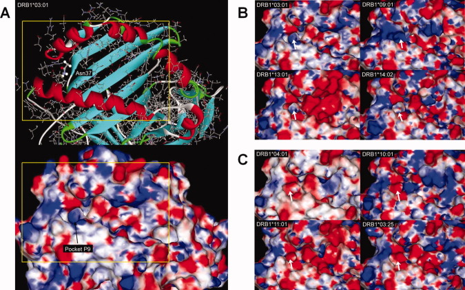
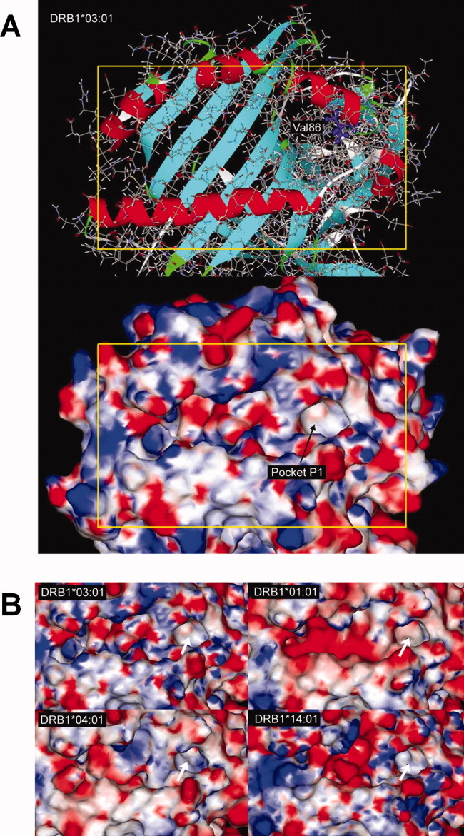
The strongest genetic risk factors for primary sclerosing cholangitis (PSC) are found in the human leukocyte antigen (HLA) complex at chromosome 6p21. Genes in the HLA class II region encode molecules that present antigen to T lymphocytes. Polymorphisms in these genes are associated with most autoimmune diseases, most likely because they contribute to the specificity of immune responses. The aim of this study was to analyze the structure and electrostatic properties of the peptide-binding groove of HLA-DR in relation to PSC. Thus, four-digit resolution HLA-DRB1 genotyping was performed in 356 PSC patients and 366 healthy controls. Sequence information was used to assign which amino acids were encoded at all polymorphic positions. In stepwise logistic regressions, variations at residues 37 and 86 were independently associated with PSC (P = 1.2 × 10(-32) and P = 1.8 × 10(-22) in single-residue models, respectively). Three-dimensional modeling was performed to explore the effect of these key residues on the HLA-DR molecule. This analysis indicated that residue 37 was a major determinant of the electrostatic properties of pocket P9 of the peptide-binding groove. Asparagine at residue 37, which was associated with PSC, induced a positive charge in pocket P9. Tyrosine, which protected against PSC, induced a negative charge in this pocket. Consistent with the statistical observations, variation at residue 86 also indirectly influenced the electrostatic properties of this pocket. DRB1*13:01, which was PSC-associated, had a positive P9 pocket and DRB1*13:02, protective against PSC, had a negative P9 pocket.
Conclusion: The results suggest that in patients with PSC, residues 37 and 86 of the HLA-DRβ chain critically influence the electrostatic properties of pocket P9 and thereby the range of peptides presented.
Copyright © 2011 American Association for the Study of Liver Diseases.
Figures

Comment in
-
Electrostatic modifications of the human leukocyte antigen DR P9 peptide-binding pocket in primary sclerosing cholangitis: back to the future with human leukocyte antigen DRβ.Hepatology. 2011 Jun;53(6):1798-800. doi: 10.1002/hep.24389. Hepatology. 2011. PMID: 21538433 No abstract available.
Similar articles
-
Electrostatic modifications of the human leukocyte antigen DR P9 peptide-binding pocket in primary sclerosing cholangitis: back to the future with human leukocyte antigen DRβ.Hepatology. 2011 Jun;53(6):1798-800. doi: 10.1002/hep.24389. Hepatology. 2011. PMID: 21538433 No abstract available.
-
Evaluation of the role of MHC class II alleles, haplotypes and selected amino acid sequences in primary sclerosing cholangitis.Autoimmunity. 2002 Dec;35(8):555-64. doi: 10.1080/0891693021000054093. Autoimmunity. 2002. PMID: 12765483
-
Genetic associations in Italian primary sclerosing cholangitis: heterogeneity across Europe defines a critical role for HLA-C.J Hepatol. 2010 May;52(5):712-7. doi: 10.1016/j.jhep.2009.11.029. Epub 2010 Mar 4. J Hepatol. 2010. PMID: 20347497
-
HLA, gut microbiome and hepatic autoimmunity.Front Immunol. 2022 Aug 18;13:980768. doi: 10.3389/fimmu.2022.980768. eCollection 2022. Front Immunol. 2022. PMID: 36059527 Free PMC article. Review.
-
The molecular genetics of autoimmune liver disease.Hepatology. 1994 Jul;20(1 Pt 1):225-39. doi: 10.1016/0270-9139(94)90157-0. Hepatology. 1994. PMID: 8020893 Review.
Cited by
-
Small duct primary sclerosing cholangitis without inflammatory bowel disease is genetically different from large duct disease.Liver Int. 2014 Nov;34(10):1488-95. doi: 10.1111/liv.12492. Epub 2014 Mar 7. Liver Int. 2014. PMID: 24517468 Free PMC article.
-
Dense genotyping of immune-related disease regions identifies nine new risk loci for primary sclerosing cholangitis.Nat Genet. 2013 Jun;45(6):670-5. doi: 10.1038/ng.2616. Epub 2013 Apr 21. Nat Genet. 2013. PMID: 23603763 Free PMC article.
-
Primary Biliary Cholangitis and Primary Sclerosing Cholangitis: Current Knowledge of Pathogenesis and Therapeutics.Biomedicines. 2022 May 31;10(6):1288. doi: 10.3390/biomedicines10061288. Biomedicines. 2022. PMID: 35740310 Free PMC article. Review.
-
Genetics in PSC: what do the "risk genes" teach us?Clin Rev Allergy Immunol. 2015 Jun;48(2-3):154-64. doi: 10.1007/s12016-014-8417-z. Clin Rev Allergy Immunol. 2015. PMID: 24736995 Review.
-
Genetics of primary sclerosing cholangitis and pathophysiological implications.Nat Rev Gastroenterol Hepatol. 2017 May;14(5):279-295. doi: 10.1038/nrgastro.2016.154. Epub 2017 Mar 15. Nat Rev Gastroenterol Hepatol. 2017. PMID: 28293027 Review.
References
-
- Bergquist A, Montgomery SM, Bahmanyar S, Olsson R, Danielsson A, Lindgren S, et al. Increased risk of primary sclerosing cholangitis and ulcerative colitis in first-degree relatives of patients with primary sclerosing cholangitis. Clin Gastroenterol Hepatol. 2008;6:939–943. - PubMed
-
- Karlsen TH, Franke A, Melum E, Kaser A, Hov JR, Balschun T, et al. Genome-wide association analysis in primary sclerosing cholangitis. Gastroenterology. 2010;138:1102–1111. - PubMed
-
- Donaldson PT, Norris S. Evaluation of the role of MHC class II alleles, haplotypes and selected amino acid sequences in primary sclerosing cholangitis. Autoimmunity. 2002;35:555–564. - PubMed
-
- Spurkland A, Saarinen S, Boberg KM, Mitchell S, Broome U, Caballeria L, et al. HLA class II haplotypes in primary sclerosing cholangitis patients from five European populations. Tissue Antigens. 1999;53:459–469. - PubMed
-
- Schrumpf E, Fausa O, Forre O, Dobloug JH, Ritland S, Thorsby E. HLA antigens and immunoregulatory T cells in ulcerative colitis associated with hepatobiliary disease. Scand J Gastroenterol. 1982;17:187–191. - PubMed
Publication types
MeSH terms
Substances
Grants and funding
LinkOut - more resources
Full Text Sources
Other Literature Sources
Research Materials