

Role for Nox1 NADPH oxidase in atherosclerosis
- PMID: 21411092
- PMCID: PMC3110523
- DOI: 10.1016/j.atherosclerosis.2011.02.028
Role for Nox1 NADPH oxidase in atherosclerosis
Abstract
Objective: Examine the contribution of Nox1 NADPH oxidase to atherogenesis.
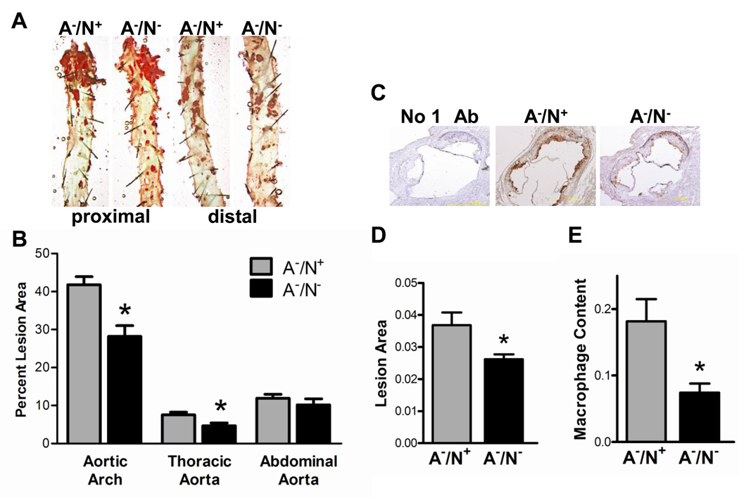
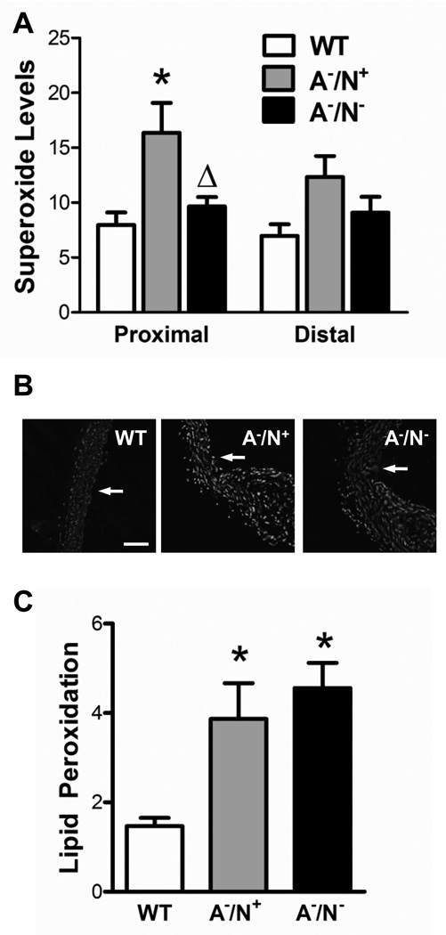
Methods and results: Male apolipoprotein E deficient mice (ApoE(-/-)) and male mice deficient in both apolipoprotein E and Nox1 (ApoE(-/-) Nox1(-/y)) received an atherogenic diet for 18 weeks. Mean blood pressures, body weights, and serum cholesterol levels were similar between the two groups of mice. Deficiency of Nox1 decreased superoxide levels and reduced lesion area in the aortic arch from 43% (ApoE(-/-)) to 28% (ApoE(-/-) Nox1(-/y)). The reduction in lesion size at the level of the aortic valve in ApoE(-/-)/Nox1(-/y) was accompanied by a decrease in macrophage infiltration as compared to ApoE(-/-) mice. Carotid artery ligation in ApoE(-/-) mice induced accelerated intimal hyperplasia with decreased cellular proliferation and increased collagen content in the neointima of vessels deficient in Nox1.
Conclusions: Nox1-derived ROS modify lesion composition and contribute to lesion size in a murine model of atherosclerosis.
Published by Elsevier Ireland Ltd.
Figures


Similar articles
-
NOX1 deficiency in apolipoprotein E-knockout mice is associated with elevated plasma lipids and enhanced atherosclerosis.Free Radic Res. 2015 Feb;49(2):186-98. doi: 10.3109/10715762.2014.992893. Free Radic Res. 2015. PMID: 25496431
-
Endothelium-restricted endothelin-1 overexpression in type 1 diabetes worsens atherosclerosis and immune cell infiltration via NOX1.Cardiovasc Res. 2021 Mar 21;117(4):1144-1153. doi: 10.1093/cvr/cvaa168. Cardiovasc Res. 2021. PMID: 32533834 Free PMC article.
-
Differential effects of NOX4 and NOX1 on immune cell-mediated inflammation in the aortic sinus of diabetic ApoE-/- mice.Clin Sci (Lond). 2016 Aug 1;130(15):1363-74. doi: 10.1042/CS20160249. Epub 2016 May 17. Clin Sci (Lond). 2016. PMID: 27190136
-
Elevated systemic TGF-beta impairs aortic vasomotor function through activation of NADPH oxidase-driven superoxide production and leads to hypertension, myocardial remodeling, and increased plaque formation in apoE(-/-) mice.Am J Physiol Heart Circ Physiol. 2010 Aug;299(2):H386-95. doi: 10.1152/ajpheart.01042.2009. Epub 2010 May 28. Am J Physiol Heart Circ Physiol. 2010. PMID: 20511416 Free PMC article.
-
NADPH oxidase 1 plays a key role in diabetes mellitus-accelerated atherosclerosis.Circulation. 2013 May 7;127(18):1888-902. doi: 10.1161/CIRCULATIONAHA.112.132159. Epub 2013 Apr 5. Circulation. 2013. PMID: 23564668
Cited by
-
8-Hydroxy-2-deoxyguanosine prevents plaque formation and inhibits vascular smooth muscle cell activation through Rac1 inactivation.Free Radic Biol Med. 2012 Jul 1;53(1):109-21. doi: 10.1016/j.freeradbiomed.2012.03.023. Epub 2012 Apr 19. Free Radic Biol Med. 2012. PMID: 22580124 Free PMC article.
-
Activation of NADPH oxidase 1 increases intracellular calcium and migration of smooth muscle cells.Hypertension. 2011 Sep;58(3):446-53. doi: 10.1161/HYPERTENSIONAHA.111.177006. Epub 2011 Aug 1. Hypertension. 2011. PMID: 21810651 Free PMC article.
-
NADPH Oxidases: From Molecular Mechanisms to Current Inhibitors.J Med Chem. 2023 Sep 14;66(17):11632-11655. doi: 10.1021/acs.jmedchem.3c00770. Epub 2023 Aug 31. J Med Chem. 2023. PMID: 37650225 Free PMC article. Review.
-
Current status of NADPH oxidase research in cardiovascular pharmacology.Vasc Health Risk Manag. 2013;9:401-28. doi: 10.2147/VHRM.S33053. Epub 2013 Jul 25. Vasc Health Risk Manag. 2013. PMID: 23983473 Free PMC article. Review.
-
NADPH oxidases: key modulators in aging and age-related cardiovascular diseases?Clin Sci (Lond). 2016 Mar;130(5):317-35. doi: 10.1042/CS20150087. Clin Sci (Lond). 2016. PMID: 26814203 Free PMC article. Review.
References
-
- Cave AC, Brewer AC, Narayanapanicker A, Ray R, Grieve DJ, Walker S, Shah AM. NADPH oxidases in cardiovascular health and disease. Antioxid Redox Signal. 2006;8:691–728. - PubMed
-
- Chlopicki S, Olszanecki R, Janiszewski M, Laurindo FR, Panz T, Miedzobrodzki J. Functional role of NADPH oxidase in activation of platelets. Antioxid Redox Signal. 2004;6:691–698. - PubMed
-
- BelAiba RS, Djordjevic T, Petry A, Diemer K, Bonello S, Banfi B, Hess J, Pogrebniak A, Bickel C, Görlach A. NOX5 variants are functionally active in endothelial cells. Free Radical Biology and Medicine. 2007;42:446–459. - PubMed
Publication types
MeSH terms
Substances
Grants and funding
LinkOut - more resources
Full Text Sources
Medical
Miscellaneous
