Endogenous MHC-related protein 1 is transiently expressed on the plasma membrane in a conformation that activates mucosal-associated invariant T cells
- PMID: 21402896
- PMCID: PMC3618670
- DOI: 10.4049/jimmunol.1003254
Endogenous MHC-related protein 1 is transiently expressed on the plasma membrane in a conformation that activates mucosal-associated invariant T cells
Abstract
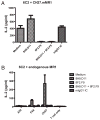
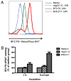
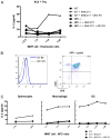
The development of mucosal-associated invariant T (MAIT) cells is dependent upon the class Ib molecule MHC-related protein 1 (MR1), commensal bacteria, and a thymus. Furthermore, recent studies have implicated MR1 presentation to MAIT cells in bacteria recognition, although the mechanism remains undefined. Surprisingly, however, surface expression of MR1 has been difficult to detect serologically, despite ubiquitous detection of MR1 transcripts and intracellular protein. In this article, we define a unique mAb capable of stabilizing endogenous mouse MR1 at the cell surface, resulting in enhanced mouse MAIT cell activation. Our results demonstrated that under basal conditions, endogenous MR1 transiently visits the cell surface, thus reconciling the aforementioned serologic and functional studies. Furthermore, using this approach, double-positive thymocytes, macrophages, and dendritic cells were identified as potential APCs for MAIT cell development and activation. Based on this pattern of MR1 expression, it is intriguing to speculate that constitutive expression of MR1 may be detrimental for maintenance of immune homeostasis in the gut and/or detection of pathogenic bacteria in mucosal tissues.
Conflict of interest statement
The authors have no financial conflicts of interest.
Figures


Similar articles
-
Evidence for MR1 antigen presentation to mucosal-associated invariant T cells.J Biol Chem. 2005 Jun 3;280(22):21183-93. doi: 10.1074/jbc.M501087200. Epub 2005 Mar 31. J Biol Chem. 2005. PMID: 15802267
-
MR1 antigen presentation to mucosal-associated invariant T cells was highly conserved in evolution.Proc Natl Acad Sci U S A. 2009 May 19;106(20):8290-5. doi: 10.1073/pnas.0903196106. Epub 2009 Apr 30. Proc Natl Acad Sci U S A. 2009. PMID: 19416870 Free PMC article.
-
A molecular basis underpinning the T cell receptor heterogeneity of mucosal-associated invariant T cells.J Exp Med. 2014 Jul 28;211(8):1585-600. doi: 10.1084/jem.20140484. Epub 2014 Jul 21. J Exp Med. 2014. PMID: 25049336 Free PMC article.
-
Expression and trafficking of MR1.Immunology. 2017 Jul;151(3):270-279. doi: 10.1111/imm.12744. Epub 2017 May 18. Immunology. 2017. PMID: 28419492 Free PMC article. Review.
-
MR1-restricted mucosal associated invariant T (MAIT) cells in the immune response to Mycobacterium tuberculosis.Immunol Rev. 2015 Mar;264(1):154-66. doi: 10.1111/imr.12271. Immunol Rev. 2015. PMID: 25703558 Free PMC article. Review.
Cited by
-
Polyclonal mucosa-associated invariant T cells have unique innate functions in bacterial infection.Infect Immun. 2012 Sep;80(9):3256-67. doi: 10.1128/IAI.00279-12. Epub 2012 Jul 9. Infect Immun. 2012. PMID: 22778103 Free PMC article.
-
Regulation of Lipid Specific and Vitamin Specific Non-MHC Restricted T Cells by Antigen Presenting Cells and Their Therapeutic Potentials.Front Immunol. 2015 Jul 28;6:388. doi: 10.3389/fimmu.2015.00388. eCollection 2015. Front Immunol. 2015. PMID: 26284072 Free PMC article. Review.
-
Distinct activation thresholds of human conventional and innate-like memory T cells.JCI Insight. 2016 Jun 2;1(8):e86292. doi: 10.1172/jci.insight.86292. JCI Insight. 2016. PMID: 27331143 Free PMC article.
-
Contribution of APCs to mucosal-associated invariant T cell activation in infectious disease and cancer.Innate Immun. 2018 May;24(4):192-202. doi: 10.1177/1753425918768695. Epub 2018 Apr 9. Innate Immun. 2018. PMID: 29631470 Free PMC article. Review.
-
MR1-dependent antigen presentation.Semin Cell Dev Biol. 2018 Dec;84:58-64. doi: 10.1016/j.semcdb.2017.11.028. Semin Cell Dev Biol. 2018. PMID: 30449535 Free PMC article. Review.
References
-
- Hashimoto K, Hirai M, Kurosawa Y. A gene outside the human MHC related to classical HLA class I genes. Science. 1995;269:693–695. - PubMed
-
- Riegert P, Wanner V, Bahram S. Genomics, isoforms, expression, and phylogeny of the MHC class I-related MR1 gene. J Immunol. 1998;161:4066–4077. - PubMed
-
- Parra-Cuadrado JF, Navarro P, Mirones I, Setién F, Oteo M, Martínez-Naves E. A study on the polymorphism of human MHC class I-related MR1 gene and identification of an MR1-like pseudogene. Tissue Antigens. 2000;56:170–172. - PubMed
-
- Yamaguchi H, Hirai M, Kurosawa Y, Hashimoto K. A highly conserved major histocompatibility complex class I-related gene in mammals. Biochem Biophys Res Commun. 1997;238:697–702. - PubMed
-
- Yamaguchi H, Kurosawa Y, Hashimoto K. Expanded genomic organization of conserved mammalian MHC class I-related genes, human MR1 and its murine ortholog. Biochem Biophys Res Commun. 1998;250:558–564. - PubMed
Publication types
MeSH terms
Substances
Grants and funding
LinkOut - more resources
Full Text Sources
Other Literature Sources
Molecular Biology Databases
Research Materials
Miscellaneous

