. 2011 Apr;138(7):1433-44.
doi: 10.1242/dev.058016.

Conditional ablation of Pten in osteoprogenitors stimulates FGF signaling

Affiliations

Conditional ablation of Pten in osteoprogenitors stimulates FGF signaling

Anyonya R Guntur et al. Development. 2011 Apr.

Abstract

Phosphatase and tensin homolog deleted on chromosome ten (PTEN) is a direct antagonist of phosphatidylinositol 3 kinase. Pten is a well recognized tumor suppressor and is one of the most commonly mutated genes in human malignancies. More recent studies of development and stem cell behavior have shown that PTEN regulates the growth and differentiation of progenitor cells. Significantly, PTEN is found in osteoprogenitor cells that give rise to bone-forming osteoblasts; however, the role of PTEN in bone development is incompletely understood. To define how PTEN functions in osteoprogenitors during bone development, we conditionally deleted Pten in mice using the cre-deleter strain Dermo1cre, which targets undifferentiated mesenchyme destined to form bone. Deletion of Pten in osteoprogenitor cells led to increased numbers of osteoblasts and expanded bone matrix. Significantly, osteoblast development and synthesis of osteoid in the nascent bone collar was uncoupled from the usual tight linkage to chondrocyte differentiation in the epiphyseal growth plate. The expansion of osteoblasts and osteoprogenitors was found to be due to augmented FGF signaling as evidenced by (1) increased expression of FGF18, a potent osteoblast mitogen, and (2) decreased expression of SPRY2, a repressor of FGF signaling. The differentiation of osteoblasts was autonomous from the growth plate chondrocytes and was correlated with an increase in the protein levels of GLI2, a transcription factor that is a major mediator of hedgehog signaling. We provide evidence that increased GLI2 activity is also a consequence of increased FGF signaling through downstream events requiring mitogen-activated protein kinases. To test whether FGF signaling is required for the effects of Pten deletion, we deleted one allele of fibroblast growth factor receptor 2 (FGFR2). Significantly, deletion of FGFR2 caused a partial rescue of the Pten-null phenotype. This study identifies activated FGF signaling as the major mediator of Pten deletion in osteoprogenitors.


Figures

Fig. 1.
Fig. 1.
Knockout of Pten using Dermo1cre. (A) Immunofluorescence images showing the loss of PTEN in both chondrocytes and osteoblasts (white boxes). The top panel shows wild-type (wt) tibial sections and the bottom panel shows those of the conditional knockout (cko) mouse. The right-hand panel shows the concomitant increase in pAKTSer473 (blue arrows) following PTEN deletion using Dermo1cre. Differential interference contrast (DIC) images are shown on the left. (B) Relative mRNA levels from wt and Dermo1cre Pten cko calvarial osteoblasts isolated from newborn pups (n=3, *P<0.05). Error bars indicate s.d.
Fig. 2.
Fig. 2.
Gross skeletal morphology, histology and microCT analysis. (A) Wild-type (wt) and conditional knockout (cko) Alizarin red- and Alcian blue-stained mouse hindlimbs showing the thicker tibia and femur (black arrows) in the Dermo1cre cko compared with those in the wt control. (B,C) Hematoxylin and Eosin (H&E) staining on 16.5 days post coitum (dpc) and one-day-old tibial sections showing increased osteoid formation in the perichondrial region (black arrows) in the cko compared with the wt control. (D) Von Kossa staining on newborn undecalcified tibial sections showing increased mineralization in the perichondrium of the Pten knockout (black arrow). (E) Bone mineral density (BMD) determined by 27 μm microCT imaging of femurs from 1-day-old wt and cko showing a significant increase in BMD (n=3, *P<0.005). Error bars indicate s.d. (F) Representative 27 μm microCT femur reconstruction data used to calculate the BMD and lengths of the femurs shown as bar graphs in E and G. (G) Length of femurs from 1-day-old wt and cko showing a significant decrease in length (n=3, *P<0.05). Error bars indicate s.d.
Fig. 3.
Fig. 3.
Cell proliferation studies using BrdU labeling. (A) Immunohistochemistry for BrdU labeled cells (dark brown) using a BrdU antibody on paraffin-embedded tissue sections. The arrow indicates the border of the perichondrium region. (B) The number of BrdU-labeled and total cells in the perichondrium (the region adjacent to the growth plate) of Pten conditional knockout (cko) and wild-type (wt) tibiae were counted and plotted as the percentage of BrdU labeled cells. A significant increase in cell proliferation was observed in the perichondrium of the cko relative to wt (n=3, *P<0.005). Error bars indicate s.d.
Fig. 4.
Fig. 4.
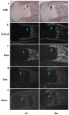
Expression of osteoblast markers in wild-type (wt) and Pten conditional knockout (cko) mice. (A) Hematoxylin and Eosin (H&E) staining on newborn tibial sections. Black arrows indicate the perichondrium region. (B) In situ hybridization on tibial sections using a Col1a1-radiolabeled probe showed an increase in transcript level in the cko relative to wt. (C-E) Transcript levels of the osteoblast differentiation markers osteopontin (Opn; C) and osterix (Osx; D) were substantially increased in the cko compared with the wt in both the perichondrium (blue arrows indicate the perichondrium) and the primary ossification region, whereas there was no difference in the expression pattern of Pthr1 (E).
Fig. 5.
Fig. 5.
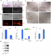
Effect of Pten deletion on FGF signaling. (A) In situ hybridization on Pten-deleted newborn tibial sections shows increased Fgf18 transcript levels in the perichondrium of Pten conditional knockout (cko) mice compared with wild type (wt), as can be observed in the dark field (DF) images. The perichondrial region is indicated with a black arrow in the H&E panel. (B) Immunofluorescence of FGF18 reveals a substantial increase in expression in the periosteum of the Pten cko compared with the wt. Black lines indicate growth plate in wt and the perichondrium in the cko. (C) Immunohistochemistry for SPRY2 expression shows decreased protein levels in the perichondrium (black arrow) of cko mice compared with wt. Black line indicates the perichondrium. (D) Overexpression of FOXO3A (FoxO3AAA) in C3H10T1/2 cells resulted in suppression of Fgf18 expression (n=3, *P<0.05). (D′) Foxo3A also inhibited the FGF18 promoter luciferase expression, which is activated by RUNX2 (Runx2) and beta-catenin (Bcat). Luciferase activity was normalized to β-galactosidase activity as shown by a co-transfected plasmid (repeated at least three times). (E) shRNAi-mediated knockdown of Pten (shRNAiPTEN) in C3H10T1/2 cells showed an increase in FGF18 transcript levels when compared with scrambled control (shRNAicntrl) (n=3, *P<0.05). (E′) Western blot showing that shRNAi can specifically knockdown PTEN protein expression and activate AKT in MC3T3E1 cells. All error bars indicate s.d. DIC, differential interference contrast.
Fig. 6.
Fig. 6.
Metatarsal rudiments isolated from the hindlimbs of wild-type (wt) and Pten conditional knockout (cko) mice at 15.5 dpc and cultured in vitro to study the cell-autonomous effect of the Pten knockout. (A) Immunofluorescence for PTEN on metatarsal rudiments showing loss of PTEN protein in the cko. (B) Hematoxylin and Eosin (H&E)-stained metatarsal rudiments showing no difference in osteoblast differentiation in the absence of PTEN (black arrows indicate osteoid). (C) Col1a1 in situ hybridization showing no change in the expression of Col1a1 mRNA. Black arrows indicate the perichondrium. Insets are low power images of the rudiments. DIC, differential interference contrast.
Fig. 7.
Fig. 7.
Effect of Pten deletion on hedgehog signaling. (A) Immunofluorescence of GLI2 showed an increase in the expression of osteoblasts that line the perichondrium in the Pten conditional knockout (cko) compared with wild type (wt). (B) In situ hybridization for patched (Ptch1), a transcriptional target for GLI2, showed an increase (black arrow) in Ptch1 expression in the cko compared with the wt control. (C,C′) Using a Gli2 luciferase reporter plasmid transfected into C3H10T1/2 cells we observed that there is an activation of Gli2 transcription when an active form of FGFR (FGFR-K650E) is co-transfected with GLI2. We also observed an increase in Gli2 luciferase activity when we used an activated MEK kinase plasmid (repeated at least three times). Black wedges indicate increasing concentrations of FGFR-K650E or MEK co-transfected with Gli2. Luciferase activity was normalized to β-galactosidase activity. (D) Using Pten flox/flox calvarial osteoblasts, we used AdenoGFP (ADGFP) as control and AdenoCRE (ADCRE) to delete PTEN as can be observed in the indirect immunofluorescence images in the top panels. When PTEN is deleted we observe an increase in pERK nuclear localization. (D′) The number of cells with a nuclear pERK signal was determined revealing a significant increase in pERK (n=3, *P<0.005). Images are overlays of DAPI (blue) and rhodamine (red) staining. (E) Western blot data for pERK and total ERK from protein lysates of Pten flox/flox calvarial osteoblasts treated with AdenoCRE and AdenoGFP. All error bars indicate s.d.
Fig. 8.
Fig. 8.
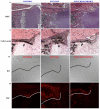
Rescue of the Pten conditional knockout (cko) mouse phenotype by deletion of one allele of Fgfr2. (A,A′) Hematoxylin and Eosin (H&E) staining of the phenotype observed in the perichondrium, along with Col1a1 in situ hybridization showing the increased perichondrium (black arrows) phenotype in the absence of Pten (PTENCKO). This phenotype is partially rescued with the deletion of Pten in the background of global loss of one allele of Fgfr2 (PTENCKO/FGFR2HET). (B,B′) Immunohistochemistry for GLI2 protein levels in the perichondrium of tibial sections showed a similar increase in GLI2 protein levels in the absence of Pten but the GLI2 levels are comparable to the wild-type levels in the Fgfr2 het Pten cko (PTENCKO/FGFR2HET) perichondrium. The conditional knockouts were generated using the Dermo1cre mouse strain. DIC, differential interference contrast.
Fig. 9.
Fig. 9.
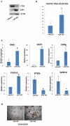
Calvarial osteoblast isolation and culture. (A) Pten flox/flox primary calvarial osteoblasts were infected with AdenoGFP (ADGFP) and AdenoCRE (ADCRE) viruses to look at knockout efficiency of Pten and downstream activation of pAKTSer473. Actin was used as a loading control. (B) In vitro proliferation assay on Pten flox/flox primary calvarial osteoblasts that were infected with AdenoGFP and AdenoCRE viruses. Osteoblasts were plated at a low density and then labeled with BrdU after allowing sufficient time for proliferation. Indirect immunofluorescence was carried out on the cells to detect nuclear BrdU; cells showing positive stain in the nucleus were counted in a field of view (n=4, *P<0.05). (C) Relative mRNA levels for the osteoblast markers were obtained from samples that were cultured in differentiation media. After deletion of Pten for 13 days (by ADCRE), osterix (OSX; n=4, *P<0.0005), osteocalcin (OCN; n=3, *P<0.005), osteopontin (OPN; n=3, *P<0.005) and FGF18 (n=3, *P<0.05) showed an increase in expression in the CRE treated samples. Pten (PTEN; n=3, *P<0.05) and Spry2 (SPRY2; n=3, *P<0.05) expression was decreased in the absence of Pten. (D) Von Kossa staining for bone nodule formation was was carried out on primary clavarial osteoblasts that were treated with the AdenoGFP and AdenoCRE viruses and cultured for 21 days in differentiation media containing β-glycerophosphate and ascorbic acid. All error bars indicate s.d.
Fig. 10.
Fig. 10.
Model of the mechanism through which PTEN regulates osteoprogenitors. Osteoblast progenitors in the perichondrium of the long bone are targeted by using Dermo1cre. When Pten is deleted in these cells, PI3K signaling effector AKT is activated. This leads to inactivation of FOXO transcriptional factors by sequestering them in the cytoplasm. This leads to downregulation of Spry2 (also known as sprouty2), a negative regulator of FGF signaling. In the Pten conditional knockout (cko), we also observe an increase in FGF18, which leads to an increase in FGF-dependent MAP Kinase activation and cell proliferation. The increase in MAP Kinase in turn activates MEK and ERK, which activate the transcription factor GLI2, leading to increased osteoblast cell differentiation.

Similar articles

Cited by

References

    1. Ambrogini E., Maria A., Marta M.-M., Ji-Hye P., Ronald A. D., Li H., Goellner J., Weinstein R. S., Jilka R. L., O'Brien C. A., et al. (2010). FoxO-mediated defense against oxidative stress in osteoblasts is indispensable for skeletal homeostasis in mice. Cell Metab. 11, 136-146 - PMC - PubMed
    1. Brewster R., Mullor J. L., Ruiz i Altaba A. (2000). Gli2 functions in FGF signaling during antero-posterior patterning. Development 127, 4395-4405 - PubMed
    1. Brunet A., Bonni A., Zigmond M. J., Lin M. Z., Juo P., Hu L. S., Anderson M. J., Arden K. C., Blenis J., Greenberg M. E. (1999). Akt promotes cell survival by phosphorylating and inhibiting a Forkhead transcription factor. Cell 96, 857-868 - PubMed
    1. Colnot C., Lu C., Hu D., Helms J. A. (2004). Distinguishing the contributions of the perichondrium, cartilage, and vascular endothelium to skeletal development. Dev. Biol. 269, 55-69 - PubMed
    1. Cooper M. K., Wassif C. A., Krakowiak P. A., Taipale J., Gong R., Kelley R. I., Porter F. D., Beachy P. A. (2003). A defective response to Hedgehog signaling in disorders of cholesterol biosynthesis. Nat. Genet. 33, 508-513 - PubMed

Publication types

MeSH terms

Substances

LinkOut - more resources