

Olmesartan, an AT1 antagonist, attenuates oxidative stress, endoplasmic reticulum stress and cardiac inflammatory mediators in rats with heart failure induced by experimental autoimmune myocarditis
- PMID: 21383952
- PMCID: PMC3048845
- DOI: 10.7150/ijbs.7.154
Olmesartan, an AT1 antagonist, attenuates oxidative stress, endoplasmic reticulum stress and cardiac inflammatory mediators in rats with heart failure induced by experimental autoimmune myocarditis
Abstract
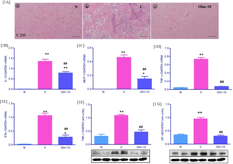
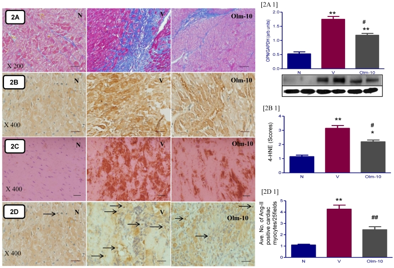
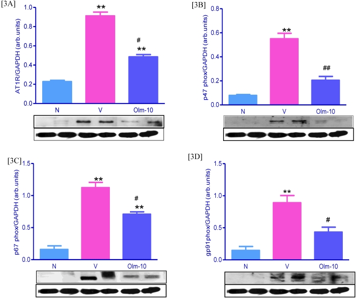
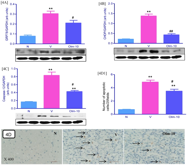
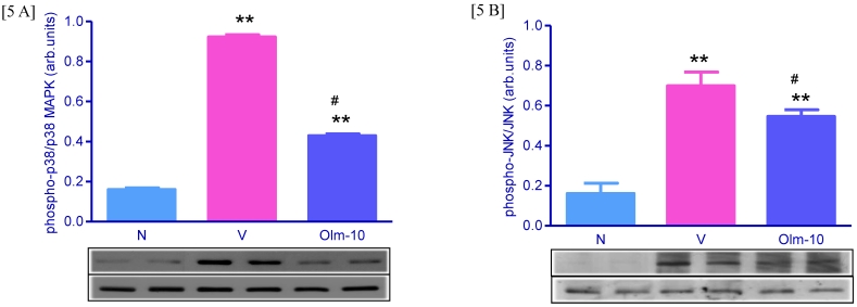
Studies have demonstrated that angiotensin II has been involved in immune and inflammatory responses which might contribute to the pathogenesis of immune-mediated diseases. Recent evidence suggests that oxidative stress may play a role in myocarditis. Here, we investigated whether olmesartan, an AT(1)R antagonist protects against experimental autoimmune myocarditis (EAM) by suppression of oxidative stress, endoplasmic reticulum (ER) stress and inflammatory cytokines. EAM was induced in Lewis rats by immunization with porcine cardiac myosin, were divided into two groups and treated with either olmesartan (10 mg/kg/day) or vehicle for a period of 21 days. Myocardial functional parameters measured by hemodynamic and echocardiographic analyses were significantly improved by the treatment with olmesartan compared with those of vehicle-treated rats. Treatment with olmesartan attenuated the myocardial mRNA expressions of proinflammatory cytokines, [Interleukin (IL)-1β, monocyte chemoattractant protein-1, tumor necrosis factor-α and interferon-γ)] and the protein expression of tumor necrosis factor-α compared with that of vehicle-treated rats. Myocardial protein expressions of AT(1)R, NADPH oxidase subunits (p47phox, p67phox, gp91phox) and the expression of markers of oxidative stress (3-nitrotyrosine and 4-hydroxy-2-nonenal), and the cardiac apoptosis were also significantly decreased by the treatment with olmesartan compared with those of vehicle-treated rats. Furthermore, olmesartan treatment down-regulated the myocardial expressions of glucose regulated protein-78, growth arrest and DNA damage-inducible gene, caspase-12, phospho-p38 mitogen-activated protein kinase (MAPK) and phospho-JNK. These findings suggest that olmesartan protects against EAM in rats, at least in part via suppression of oxidative stress, ER stress and inflammatory cytokines.
Keywords: Experimental autoimmune myocarditis; endoplasmic reticulum stress; inflammation; olmesartan; oxidative stress.
Conflict of interest statement
Conflict of Interests: The authors have declared that no conflict of interest exists.
Figures
Similar articles
-
Olmesartan attenuates the development of heart failure after experimental autoimmune myocarditis in rats through the modulation of ANG 1-7 mas receptor.Mol Cell Endocrinol. 2012 Apr 4;351(2):208-19. doi: 10.1016/j.mce.2011.12.010. Epub 2011 Dec 19. Mol Cell Endocrinol. 2012. PMID: 22200414
-
Beneficial effects of olmesartan, an angiotensin II receptor type 1 antagonist, in rats with dilated cardiomyopathy.Exp Biol Med (Maywood). 2010 Nov;235(11):1338-46. doi: 10.1258/ebm.2010.010016. Epub 2010 Sep 27. Exp Biol Med (Maywood). 2010. PMID: 20876084
-
Cardioprotective effects of telmisartan against heart failure in rats induced by experimental autoimmune myocarditis through the modulation of angiotensin-converting enzyme-2/angiotensin 1-7/mas receptor axis.Int J Biol Sci. 2011;7(8):1077-92. doi: 10.7150/ijbs.7.1077. Epub 2011 Sep 8. Int J Biol Sci. 2011. PMID: 21927577 Free PMC article.
-
Effect of olmesartan medoxomil on atherosclerosis: clinical implications of the emerging evidence.Am J Cardiovasc Drugs. 2006;6(6):363-6. doi: 10.2165/00129784-200606060-00002. Am J Cardiovasc Drugs. 2006. PMID: 17192125 Review.
-
Regulation of inflammation and myocardial fibrosis in experimental autoimmune myocarditis.Inflamm Allergy Drug Targets. 2011 Jun;10(3):218-25. doi: 10.2174/187152811795564091. Inflamm Allergy Drug Targets. 2011. PMID: 21495969 Review.
Cited by
-
Endoplasmic reticulum stress and diabetic cardiomyopathy.Exp Diabetes Res. 2012;2012:827971. doi: 10.1155/2012/827971. Epub 2011 Nov 24. Exp Diabetes Res. 2012. PMID: 22144992 Free PMC article. Review.
-
Recent Advances on Drug Development and Emerging Therapeutic Agents Through Targeting Cellular Homeostasis for Ageing and Cardiovascular Disease.Front Aging. 2022 Apr 25;3:888190. doi: 10.3389/fragi.2022.888190. eCollection 2022. Front Aging. 2022. PMID: 35821839 Free PMC article. Review.
-
Endoplasmic reticulum stress in the heart: insights into mechanisms and drug targets.Br J Pharmacol. 2018 Apr;175(8):1293-1304. doi: 10.1111/bph.13888. Epub 2017 Jun 27. Br J Pharmacol. 2018. PMID: 28548229 Free PMC article. Review.
-
Role of ER Stress Mediated Unfolded Protein Responses and ER Stress Inhibitors in the Pathogenesis of Inflammatory Bowel Disease.Dig Dis Sci. 2022 Dec;67(12):5392-5406. doi: 10.1007/s10620-022-07467-y. Epub 2022 Mar 22. Dig Dis Sci. 2022. PMID: 35318552 Review.
-
Suppression of endoplasmic reticulum stress improves endothelium-dependent contractile responses in aorta of the spontaneously hypertensive rat.Am J Physiol Heart Circ Physiol. 2013 Aug 1;305(3):H344-53. doi: 10.1152/ajpheart.00952.2012. Epub 2013 May 24. Am J Physiol Heart Circ Physiol. 2013. PMID: 23709602 Free PMC article.
References
-
- Kawai C. From myocarditis to cardiomyopathy: mechanisms of inflammation and cell death. Circulation. 1999;99:1091–1100. - PubMed
-
- Feldman AM, McNamara D. Myocarditis. N Engl J Med. 2000;343:1388–1398. - PubMed
-
- Kodama M, Matsumoto Y, Fujiwara M, Masani F, Izumi T, Shibata A. A novel experimental model of giant cell myocarditis induced in rats by immunization with cardiac myosin fraction. Clin Immunol Immunopathol. 1990;57:250–262. - PubMed
-
- Kodama M, Matsumoto Y, Fujiwara M, Zhang SS, Hanawa H, Itoh E, Tsuda T, Izumi T, Shibata A. Characteristics of giant cells and factors related to the formation of giant cells in myocarditis. Circ Res. 1991;69:1042–1050. - PubMed
-
- Kodama M, Matsumoto Y, Fujiwara M. In vivo lymphocytemediated myocardial injures demonstrated by adoptive transfer of experimental autoimmune myocarditis. Circulation. 1992;85:1918– 1926. - PubMed
Publication types
MeSH terms
Substances
LinkOut - more resources
Full Text Sources
Other Literature Sources
Medical
Research Materials
