. 2011 Apr;18(4):471-7.
doi: 10.1038/nsmb.2004. Epub 2011 Mar 6.

The structural basis for MCM2-7 helicase activation by GINS and Cdc45

Affiliations

The structural basis for MCM2-7 helicase activation by GINS and Cdc45

Alessandro Costa et al. Nat Struct Mol Biol. 2011 Apr.

Abstract

Two central steps for initiating eukaryotic DNA replication involve loading of the Mcm2-7 helicase onto double-stranded DNA and its activation by GINS-Cdc45. To better understand these events, we determined the structures of Mcm2-7 and the CMG complex by using single-particle electron microscopy. Mcm2-7 adopts two conformations--a lock-washer-shaped spiral state and a planar, gapped-ring form--in which Mcm2 and Mcm5 flank a breach in the helicase perimeter. GINS and Cdc45 bridge this gap, forming a topologically closed assembly with a large interior channel; nucleotide binding further seals off the discontinuity between Mcm2 and Mcm5, partitioning the channel into two smaller pores. Together, our data help explain how GINS and Cdc45 activate Mcm2-7, indicate that Mcm2-7 loading may be assisted by a natural predisposition of the hexamer to form open rings, and suggest a mechanism by which the CMG complex assists DNA strand separation.


Figures
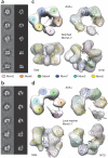
Figure 1
Figure 1
Mcm2–7 exists in two states. (a,b) Reference-free class averages and corresponding forward projections of notched-ring or lock-washer reconstructions. (c) 3D reconstruction of the notched ring viewed from the AAA+ face (full view, left; slab view, right), the side and the N-terminal face. Mcm homology models are fitted into the reconstruction. (d) 3D structure of the lock-washer ring viewed from the AAA+ (full or slab view), side and N-terminal faces, and fitted with homology models.
Figure 2
Figure 2
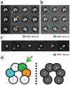
Subunit mapping of Mcm2–7. (a) Projection per class average matching of the Mcm2–7 lock-washer state with an N-terminal MBP tag on Mcm6. Orange arrowheads mark additional density observed on the ring. (b) Class averages of Mcm2–7 containing MBP-tagged Mcm3. Orientations match those in a. Blue arrowheads mark additional density observed on the ring. (c) Class-average projection matching of Mcm2–7 containing MBP-tagged Mcm4. The tag appears detached from the ring density (green arrowheads), consistent with the presence of a 154-residue N-terminal tail on Mcm4. (d) Possible subunit arrangements for gapped Mcm2–7 complexes as defined by MBP-tagged Mcm3 and Mcm6. The correct configuration further defined by MBP-tagged Mcm4 is shown in color.
Figure 3
Figure 3
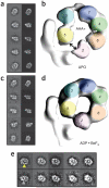
CMG contains a notched, planar Mcm2–7 ring that is sealed upon nucleotide binding. (a) Class-average projection matching for apo CMG. (b) AAA+ view of apo CMG, showing a discontinuity between Mcm2–5. (c) Class-average projection matching for ADP·BeF3-bound CMG. (d) C-terminal AAA+ view of ADP·BeF3-bound CMG, showing a pinched-off gap between Mcm2–5. (e) Class averages of apo (top) or ADP·BeF3-bound CMG (bottom) in the same orientation, showing the presence (yellow arrowhead) or absence (gray arrowhead) of an Mcm2–5 gap.
Figure 4
Figure 4
Mcm-subunit mapping in the CMG. (ac) Class-average projection matching for ADP·BeF3-bound CMG with (unboxed) or without (boxed) N-terminal MBP tags (arrowheads) on Mcm6 (a), Mcm3 (b) or Mcm4 (c). Mcm4 is distal from GINS and Cdc45 (see also Supplementary Fig. 5). (dg) Unfiltered 3D reconstructions of ADP·BeF3-bound CMG with an N-terminal MBP tag on either Mcm6 (d) or Mcm3 (e) and compared to both untagged CMG (f) and a CMG complex with a 154-amino-acid deletion of the N-terminal Mcm4 tail (g). Extra density attributed to MBP on Mcm6 and Mcm3 is colored orange or light blue, respectively; only a portion of the Mcm4 tail is observable in the full-length constructs and is highlighted in green. (h) Density corresponding to MBP on Mcm3 (light blue) or Mcm6 (orange), cut out from the CMG density maps and compared to the density map calculated from the MBP crystal structure, PDB entry 3HPI, filtered to 30 Å and showed at 5σ (gray).
Figure 5
Figure 5
Structure docking into CMG reconstructions. (a,b) From left to right: N-terminal, side and C-terminal (AAA+ domain) views of apo (a) or ADP·BeF3-bound CMG (b). The red double arrow in a indicates the location of a discontinuity in the AAA+ tier of Mcm2–7 in apo CMG. GINS is colored white; unoccupied density assigned to Cdc45 is solid gray.
Figure 6
Figure 6
GINS contacts within the CMG. (a) Crystal structures of the four individual subunits of the GINS complex. As Sld5 and Psf1 are close homologs, an orientation for the β-domain in Psf1 was generated using that seen in Sld5. The related β-domains of Psf1 and Psf3 are shown in black and highlighted with gold ovals. Psf2 and Psf3 share close structural similarity; a related helical element (α1) is shown in black and highlighted with gold ovals. (b) Three views of the crystal structure of GINS docked into the CMG density. (c) Zoomed-out (left) and close-up (right) views of GINS engaged with Cdc45 and the N-terminal domains of Mcm3 and Mcm5. (d) Zoomed-out (left) and close-up (right) views of GINS and its interactions with Cdc45 and the AAA+ domains of Mcm3 and Mcm5. (e) Nucleotide-triggered movements in the GINS-AAA+ domain interaction region appear to seal off the Mcm2–7 central channel.
Figure 7
Figure 7
CMG interactions. (a) Expression levels of individual proteins used for testing CMG formation. (b) Immunoprecipitation (IP) experiments testing CMG stability. 1, wild-type CMG; 2, CMG with a C-terminal truncation in Psf1; 3, CMG without MCM5; 4, CMG without Cdc45. Mcm3 immunoprecipitation yields the intact CMG complex, whereas deletion of the Psf1 β domain (Psf1ΔC, amino acids 1–139), or withholding Mcm5 or Cdc45, disrupts CMG formation. (c) Expression levels of individual proteins used for testing GINS formation. (d) IP experiments testing GINS stability. The C-terminal β-domain of Psf1 is dispensable for GINS formation. (e) Summary of interactions in the CMG. Subunits are demarcated by spheres, which have been placed into the 3D reconstruction obtained for the ADP·BeF3-bound complex (transparent surface). The N- and C-terminal regions of each Mcm subunit are highlighted separately and labeled 2N–7N and 2C–7C, respectively; other subunits are labeled by their full name. Dotted lines show noncovalent interactions observed in the complex and are colored as follows: black, intra-MCM; blue, intra-GINS; green, GINS with Cdc45; red, GINS and Cdc45 with Mcm2–7.
Figure 8
Figure 8
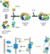
Model for Mcm2–7 activation and function. (a) Free Mcm2–7 can exist in either an open, lock-washer or notched, planar configuration. Each form shows a discontinuity between Mcm2 and Mcm5. Binding of GINS–Cdc45 stabilizes the notched, planar Mcm2–7 state, whereas ATP binding promotes ring closure. (b) ORC, Cdc6 and Cdt1 load a preopened Mcm2–7 assembly onto dsDNA as an inactive double hexamer. GINS and Cdc45 bind to Mcm2–7, concomitant with an isomerization that creates or stabilizes melted DNA. The side channel formed by the GINS–Cdc45 subcomplex likely prevents DNA escape from Mcm2–7 and may help partition the lagging DNA strand from its complement following unwinding.

Similar articles

Cited by

References

    1. Davey MJ, O’Donnell M. Replicative helicase loaders: ring breakers and ring makers. Curr. Biol. 2003;13:R594–R596. - PubMed
    1. Funnell BE, Baker TA, Kornberg A. In vitro assembly of a prepriming complex at the origin of the Escherichia coli chromosome. J. Biol. Chem. 1987;262:10327–10334. - PubMed
    1. Davey MJ, Fang L, McInerney P, Georgescu RE, O’Donnell M. The DnaC helicase loader is a dual ATP/ADP switch protein. EMBO J. 2002;21:3148–3159. - PMC - PubMed
    1. Valle M, Gruss C, Halmer L, Carazo JM, Donate LE. Large T-antigen double hexamers imaged at the simian virus 40 origin of replication. Mol. Cell. Biol. 2000;20:34–41. - PMC - PubMed
    1. Kumar A, et al. Model for T-antigen-dependent melting of the simian virus 40 core origin based on studies of the interaction of the beta-hairpin with DNA. J. Virol. 2007;81:4808–4818. - PMC - PubMed

Publication types

MeSH terms

Substances

LinkOut - more resources