Review
. 2011:40:337-59.
doi: 10.1146/annurev-biophys-042910-155338.

Protein folding at the exit tunnel

Affiliations
Review

Protein folding at the exit tunnel

Daria V Fedyukina et al. Annu Rev Biophys. 2011.

Abstract

Over five decades of research have yielded a large body of information on how purified proteins attain their native state when refolded in the test tube, starting from a chemically or thermally denatured state. Nevertheless, we still know little about how proteins fold and unfold in their natural biological habitat: the living cell. Indeed, a variety of cellular components, including molecular chaperones, the ribosome, and crowding of the intracellular medium, modulate folding mechanisms in physiologically relevant environments. This review focuses on the current state of knowledge in protein folding in the cell with emphasis on the early stage of a protein's life, as the nascent polypeptide traverses and emerges from the ribosomal tunnel. Given the vectorial nature of ribosome-assisted translation, the transient degree of chain elongation becomes a relevant variable expected to affect nascent protein foldability, aggregation propensity and extent of interaction with chaperones and the ribosome.


Figures

Figure 1
Figure 1
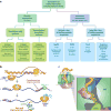
Scheme illustrating limiting in vitro protein folding mechanisms denoted by dashed gray (rarely observed), dark blue, and black arrows. The experiments leading to the formulation of these models are typically performed in purified protein solutions and involve the refolding of unfolded states generated chemically or by temperature jumps. Note that the species other than the unfolded and folded states (denoted 2, 3, and 4) may be either intermediates, transition states, or transient species populated along diffusive downhill routes.
Figure 2
Figure 2
Schematic representation of key aspects of cotranslational protein folding in the crowded milieu of the cellular cytosol.
Figure 3
Figure 3
(a) Crystal structure of the Escherichia coli ribosome at 3.5 Å resolution (PDB IDs: 2AVY and 2AW4) (72). The ribosomal RNA is represented as surfaces (23S and 5S RNAs, turquoise; 16S RNA, beige). Ribosomal proteins are shown as ribbons (proteins in 50S subunit, purple; proteins in 30S subunit, green). Schematic representation of a vertical section of the 70S (b) prokaryotic and (c) archaeal ribosomes highlighting the ribosomal proteins facing or near the exit tunnel and the ribosome-associated TF chaperone. A representative hypothetical nascent polypeptide is drawn in yellow. (d) Structure of the ribosomal exit tunnel (PDB file kindly provided by N.R. Voss and P.B. Moore) (85). Abbreviations: PTC, peptidyl transferase center; TF, trigger factor.
Figure 4
Figure 4
Model for the dynamic interaction of the trigger factor (TF) chaperone with ribosomes. The symbol t1/2 denotes the half-life for the dissociation of the TF-ribosome binary complex or the apparent half-life for the dissociation of the TF-ribosome-nascent chain ternary complex. (a,b) The apparent association rate constant of TF (green) to ribosomes increases when a peptide chain emerges from the ribosomal exit tunnel. (c) Some longer nascent chains can increase the half-life t1/2 for complex dissociation up to ~53 s. (d) The association rate of TF for ribosomes eventually decreases when a large nascent polypeptide is exposed to the ribosomal surface. TF may remain associated with some nascent chains even after dissociation of TF from its ribosome-binding site. Nonpolar stretches serving as TF binding sites are in blue. Adapted from Reference .
Figure 5
Figure 5
(a) Overview of currently available methods to generate RNCs of well-defined chain length. Step-by-step procedures based on (b) in vitro (cell-free) coupled transcription-translation and (c) SecM stalling. For simplicity, cotranslationally active chaperones are omitted. The 17-residue SecM peptide-stalling sequence (FXXXXWIXXXXGIRAGP) is shown inside the ribosomal tunnel. The underlined amino acids (in red) experience critical interactions with the ribosomal tunnel (white dashed lines) with L22. Abbreviations: RNAP, RNA polymerase; RNC, ribosome-bound nascent chain; X, any residue.
Figure 6
Figure 6
Relationship between specific RNC structural features and biological or spectroscopic techniques employed to elucidate them. Abbreviations: cryo-EM, cryo-electron microscopy; FRET, Förster resonance energy transfer; NMR, nuclear magnetic resonance; RNC, ribosome-bound nascent chain.
Figure 7
Figure 7
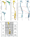
Cryo-EM maps of different peptidyl tRNAs inside the eukaryotic ribosome’s P-site and exit tunnel. (a) 80S–helix 1 RNC, (b) 80S–DPAP RNC, (c) 80S–helix2 RNC, and (d) enlarged view of transparent density of panel a with fitted ribbon model for tRNA and nascent chain. (e, f) Enlarged view of panel c with alternative models for helix 2 nascent chain. Red arrows indicate corresponding region (residues 97–108) modeled as helical (e) or extended (f). (g) Schematic cross-section of 80S–helix 1 RNC representing helix formation within the exit tunnel. Abbreviations: cryo-EM, cyro-electron microscopy; RNC, ribosome-bound nascent chain; DPAP, dipeptidylaminopeptidase; PTC, peptidyl transferase center. Adapted by permission from Macmillan Publishers Ltd: Nature Structural & Molecular Biology (Reference 5), copyright (2010).
Figure 8
Figure 8
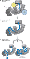
Mode for TF chaperone binding to nascent polypeptides based on cross-linking experiments by Merz et al. (59). TF directs the nascent chains through its interior in a sequence- and length-dependent manner. Interactions with TF are (a) moderate for nascent chains 40 to 60 residues long, and (b) considerable for nascent chains up to 90 residues, where the nascent chain’s N terminus reaches up to the TF PPIase domain (head). (c) Upon further elongation, the nascent chain may leave TF or it may accumulate in the interior of the TF chaperone. Abbreviations: PDF, protein deformylase; MAP, methionine aminopeptidase; SRP, signal recognition particle; TF, trigger factor. Adapted by permission from Macmillan Publishers Ltd: EMBO Journal (Reference 59), copyright (2008).
Figure 9
Figure 9
(a) Frequency domain dynamic fluorescence depolarization of ribosome-bound apoMb and PIR nascent chains generated in an Escherichia coli cell-free system. Data are shown only for the nanosecond local motions that reveal the presence of a small compact or semicompact species. (b) Scheme highlighting the motions associated with each fluorescence phase with each associated component of the motion. (c) Scheme illustrating the spatial amplitude of the subnanosecond local motion of the N terminus of the fluorophore-labeled RNC. The symbol θo represents the cone semiangle (in red) assessed in panel d. (d) Amplitude of the fast (subnanosecond) motions experienced by the N termini of nascent apoMb and natively unfolded PIR nascent polypeptides of increasing length under different conditions. Data were collected for samples prepared from either wild-type or Δtig TF-depleted cell strains. Panels a and b adapted with permission from References and , respectively. Copyright 2008 and 2009, respectively, American Chemical Society and John Wiley and Sons. Abbreviations: apoMb, apomyoglobin; PIR, phosphorylated insulin receptor interaction region; RNC, ribosome-bound nascent chain; TF, trigger factor chaperone.
Figure 10
Figure 10
Model for the cotranslational folding of P22 tailspike nascent protein chains. Abbreviations: TSS, tailspike short stalled nascent chain; TMS, tailspike mid-length stalled nascent chain; TβS, tailspike stalled nascent chain with the entire β-helix exposed; TFS, tailspike full stalled nascent chain. Reprinted from the Journal of Molecular Biology, Vol. 383, Evans MS, Sander IM, Clark PL. “Cotranslational folding promotes beta-helix formation and avoids aggregation in vivo” pp. 683–92, Copyright (2008), with permission from Elsevier.

Similar articles

Cited by

References

    1. Anfinsen CB, Redfield RR, Choate WI, Page J, Carroll WR. Studies on the gross structure, cross-linkages, and terminal sequences in ribonuclease. J Biol Chem. 1954;207:201–10. - PubMed
    1. Ban N, Nissen P, Hansen J, Moore PB, Steitz TA. The complete atomic structure of the large ribosomal subunit at 2.4 angstrom resolution. Science. 2000;289:905–20. - PubMed
    1. Baram D, Pyetan E, Sittner A, Auerbach-Nevo T, Bashan A, Yonath A. Structure of trigger factor binding domain in biologically homologous complex with eubacterial ribosome reveals its chaperone action. Proc Natl Acad Sci USA. 2005;102:12017–22. - PMC - PubMed
    1. Batey S, Nickson AA, Clarke J. Studying the folding of multidomain proteins. HFSP J. 2008;2:365–77. - PMC - PubMed
    1. Bhushan S, Gartmann M, Halic M, Armache J-P, Jarasch A, et al. Alpha-helical nascent polypeptide chains visualized within distinct regions of the ribosomal exit tunnel. Nat Struct Mol Biol. 2010;17:313–17. - PubMed

Publication types

LinkOut - more resources