. 2011 May;188(1):105-25.
doi: 10.1534/genetics.111.127910. Epub 2011 Mar 2.

Identification of novel Ras-cooperating oncogenes in Drosophila melanogaster: a RhoGEF/Rho-family/JNK pathway is a central driver of tumorigenesis

Affiliations

Identification of novel Ras-cooperating oncogenes in Drosophila melanogaster: a RhoGEF/Rho-family/JNK pathway is a central driver of tumorigenesis

Anthony M Brumby et al. Genetics. 2011 May.

Abstract

We have shown previously that mutations in the apico-basal cell polarity regulators cooperate with oncogenic Ras (Ras(ACT)) to promote tumorigenesis in Drosophila melanogaster and mammalian cells. To identify novel genes that cooperate with Ras(ACT) in tumorigenesis, we carried out a genome-wide screen for genes that when overexpressed throughout the developing Drosophila eye enhance Ras(ACT)-driven hyperplasia. Ras(ACT)-cooperating genes identified were Rac1 Rho1, RhoGEF2, pbl, rib, and east, which encode cell morphology regulators. In a clonal setting, which reveals genes conferring a competitive advantage over wild-type cells, only Rac1, an activated allele of Rho1 (Rho1(ACT)), RhoGEF2, and pbl cooperated with Ras(ACT), resulting in reduced differentiation and large invasive tumors. Expression of RhoGEF2 or Rac1 with Ras(ACT) upregulated Jun kinase (JNK) activity, and JNK upregulation was essential for cooperation. However, in the whole-tissue system, upregulation of JNK alone was not sufficient for cooperation with Ras(ACT), while in the clonal setting, JNK upregulation was sufficient for Ras(ACT)-mediated tumorigenesis. JNK upregulation was also sufficient to confer invasive growth of Ras(V12)-expressing mammalian MCF10A breast epithelial cells. Consistent with this, HER2(+) human breast cancers (where human epidermal growth factor 2 is overexpressed and Ras signaling upregulated) show a significant correlation with a signature representing JNK pathway activation. Moreover, our genetic analysis in Drosophila revealed that Rho1 and Rac are important for the cooperation of RhoGEF2 or Pbl overexpression and of mutants in polarity regulators, Dlg and aPKC, with Ras(ACT) in the whole-tissue context. Collectively our analysis reveals the importance of the RhoGEF/Rho-family/JNK pathway in cooperative tumorigenesis with Ras(ACT).


Figures
F<sc>igure</sc> 1.—
Figure 1.—
- Strategy for RasACT-cooperating gene screen: scanning electron micrographs of adult eyes (dorsal and lateral views) from female or males flies expressing oncogenic Ras (RasACT) and activated aPKC (aPKCΔN) or knockdown of Dlg (dlgRNAi), compared with RasACT alone and controls. Posterior is to the left and dorsal is to the top in the lateral views, in this and all other adult eye figures. (A) ey-GAL4 UAS-GFP (ey>GFP), (B) ey-GAL4 UAS-RasACT (ey>RasACT), (C) ey-GAL4 UAS-dlgRNAi (ey>dlgRNAi), (D) ey-GAL4 UAS-RasACT UAS-dlgRNAi (ey>RasACT dlgRNAi), (E) ey-GAL4 UAS-aPKCΔN (ey>aPKCΔN), (F) ey-GAL4 UAS-RasACT aPKCΔN (ey>RasACT aPKCΔN). RasACT expression via the ey-GAL4 driver results in hyperplastic eyes. Expression of aPKCΔN or dlgRNAi via ey-GAL4 results in slight roughening. Expression of aPKCΔN or dlgRNAi with RasACT via ey-GAL4 results in enhanced overgrowth of the RasACT hyperplastic adult eye. Males expressing RasACT and aPKCΔN die at the pupal stage. (G) Diagram of ey>RasACT screening strategy. ey-GAL4 UAS-RasACT flies were crossed to a library of enhancer P (GS) lines, expressing via UAS(GAL4) adjacent genes (UAS-gene “X”), and those which enhanced the ey>RasACT phenotype were selected for further analysis.
F<sc>igure</sc> 2.—
Figure 2.—
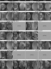
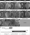
- Interaction of cooperating GS lines with ey>RasACT and validation: scanning electron micrographs of adult eyes (dorsal and lateral views) from female or male flies expressing RasACT and cooperating GS lines or UAS transgenes via the ey driver, compared with ey>RasACT alone and wild-type control eyes. (A) Control (w1118), (B) ey>RasACT, (C) ey>RasACT GS13019 (Rac1), (D) ey>RasACT Rac1, (E) ey>RasACT GS12503 (Rho1), (F) ey>RasACT Rho1ACT, (G) ey>RasACT GS45 (RhoGEF2), (H) ey>RasACT RhoGEF2, (I) ey>RasACT GS14458 (pbl), (J) ey>RasACT pblGFP#8, (K) ey>RasACT GS9641 (rib), (L) ey>RasACT GS1211 (east), (M) ey>RasACT east. Expression of ey>RasACT with GS9641 (rib), GS12503 (Rho1), or RhoGEF2 was male lethal, and with rib was male and female lethal.
F<sc>igure</sc> 3.—
Figure 3.—
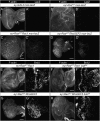
- The JNK pathway is upregulated in ey>RasACT + Rac1 or ey>RasACT + RhoGEF2, and blocking JNK reduces the overgrowth by decreasing the number of S phases: (A–D) LacZ staining of eye discs from female larvae of: (A) ey-GAL4 msn-lacZ, (B) ey>RasACT msn-lacZ, (C) ey>RasACT Rac1 msn-lacZ, (D) ey>RasACT RhoGEF2 msn-lacZ. Rac1 or RhoGEF2-expressing eye discs show increased LacZ staining (msn expression). (E–H) BrdU labeling of eye discs from: (E) ey>RasACT Rac1, (F) ey>RasACT Rac1 bskDN, (G) ey>RasACT RhoGEF2, (H) ey>RasACT RhoGEF2 bskDN. The ectopic S phases are suppressed by blocking JNK signaling with bskDN. In E, the arrowhead marks the second mitotic wave. Scale bars, 50 μm.
F<sc>igure</sc> 4.—
Figure 4.—
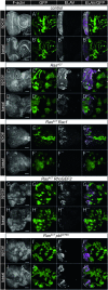
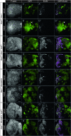
- Analysis of the cooperation of Rac1, RhoGEF2, and pbl with RasACT in eye disc clones: expression of RasACT with cooperating oncogenes in clones compared with controls from day 5 staged larvae stained for F-actin and ELAV. Clones are positively marked by GFP. (A) FRT82B control, apical section; (B) FRT82B control, basal section; (C) RasACT clones, apical section; (D) RasACT clones, basal section; (E) RasACT + Rac1, apical section; (F) RasACT + Rac1, basal section; (G) RasACT + RhoGEF2, apical section; (H) RasACT + RhoGEF2, basal section; (I) RasACT + pblGFP#3, apical section; (J) RasACT + pblGFP#3, basal section. In C, the arrowhead points to a patch of ectopic differentiation. Expression of Rac1, RhoGEF2, or pblGFP#3 with RasACT results in large overgrowths, more prominently in basal sections. Non-cell-autonomous overgrowth is also observed around the clones as evidenced by tissue folding seen by F-actin staining. Scale bars, 50 μm.
F<sc>igure</sc> 5.—
Figure 5.—
- Rac1 or RhoGEF2 expression in clones results in upregulation of the JNK pathway: LacZ staining, to detect msn-lacZ expression, and F-actin staining of mosaic discs expressing Rac1 or RhoGEF2 +/− RasACT. Clones are positively marked by GFP. (A) RasACT clones in msn-lacZ/+ discs, (B) RasACT + Rac1 clones in msn-lacZ/+ discs, apical view, (C) RasACT + Rac1 clones in msn-lacZ/+ discs, basal view, (D) RasACT + Rac1 clones in msn-lacZ/+ discs with brain lobes, showing invasion of GFP+ tissue between the brain lobes, BL, (E) RhoGEF2 clones in msn-lacZ/+ discs, (F) RasACT + RhoGEF2 clones in msn-lacZ/+ discs, apical view, (G) RasACT +RhoGEF2 clones in msn-lacZ/+ discs, basal view. In D, the arrowhead points to GFP+ tissue invading between the brain lobes. RasACT results in mild ectopic msn-lacZ expression. RasACT +RhoGEF2 and RasACT + Rac1 clones show extensive upregulation of msn-lacZ. In some RhoGEF2-expressing clones, high levels of msn-lacZ expression is observed. Scale bars, A–C, E–G, 50 μm; D, 200 μm.
F<sc>igure</sc> 6.—
Figure 6.—
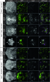
- JNK signaling is required for cooperation of Rac1 or RhoGEF2 with RasACT in clones: clones expressing RasACT with Rac1 or RhoGEF2 +/− bskDN at day 5, stained for F-actin and ELAV. Clones are positively marked by GFP. (A) RasACT + Rac1 clones, apical view; (B) RasACT + Rac1 clones, basal view; (C) RasACT + Rac1 + bskDN clones, apical view; (D) RasACT + Rac1 + bskDN clones, basal view; (E) RasACT + RhoGEF2 clones, apical view; (F) RasACT + RhoGEF2 clones, basal view; (G) RasACT + RhoGEF2 + bskDN clones, apical view; (H) RasACT + RhoGEF2 + bskDN clones, basal view. In B, the arrowhead points to tissue with an invasive morphology. Expression of bskDN rescues the cooperation of Rac1 or RhoGEF2 with RasACT by increasing differentiation and reducing the invasive phenotype of Rac1 + RasACT clones. Scale bars, 50 μm.
F<sc>igure</sc> 7.—
Figure 7.—
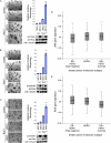
- Cooperation of JNK and Ras signaling in normal mammalian epithelial cells and human cancer: (A–C) JNK1a1, MKK4, and MKK7 cooperate with Ha-RasV12 in promoting invasion of MCF10A cells in 3D matrigel cultures. A representative of three independent experiments with three independently derived MCF10A cell line sets overexpressing JNK1a1 (A), MKK4 (B), or MKK7 (C) in the context of Ha-RasV12 expression is shown. Bright-field images of acini morphology (left) (scale bar, 100 μm). Invasive morphology quantitation expressed relative to Ha-RasV12 control [*P < 0.05; Student’s t-test, two tailed, unpaired; error bars represent standard deviation] (right). Western blot of whole-cell lysates probed with antibodies as indicated on the right side. α-Tubulin was used as loading control. (D, E) Box-plots representing expression of a gene signature of JNK activation and Ras activation in human breast cancers. The relative expression of JNK signature genes (D) and Ras signature genes (E) were compared using gene expression data and divided according to the three major molecular breast cancer subtypes: ER/HER2 (triple negative), HER2+, and ER+/HER2 (luminal). The correlation with high JNK signature expression in HER2+ and ER+/HER2 is higher than in the other breast cancer subtypes, and of these the Ras signature is higher in the HER2+ subtype (P < 0.0001).
F<sc>igure</sc> 8.—
Figure 8.—
Model of interactions of RasACT-cooperating genes: genetic epistasis tests reveal a possible model of how the RasACT-cooperating genes may function in a pathway to lead to the upregulation of JNK in the ey-GAL4 system. Expression of RasACT alone via ey-GAL4 leads to hyperplasia. Pathways shown in black lead to increased hyperplasia, while those in red lead to differentiation defects and morphological changes, which we propose is due to higher levels of Rho1 and JNK signaling. Proteins shown in green are hypothesized to be required for the effects, on the basis of information in other systems (see text). Since JNK is required, but not sufficient, to cooperate with ey>RasACT, we hypothesize that other factors (X or Y) are altered by Rho1 or Rac1 signaling to enable cooperation with RasACT. Whether knockdown of Dlg or activation of aPKC requires Pbl or RhoGEF2 is not known (indicated by ?). See text for further details.

Similar articles

Cited by

References

    1. Adachi-Yamada T., O'Connor M. B., 2004. Mechanisms for removal of developmentally abnormal cells: cell competition and morphogenetic apoptosis. J. Biochem. 136: 13–17 - PubMed
    1. Aigaki T., Ohsako T., Toba G., Seong K., Matsuo T., 2001. The gene search system: its application to functional genomics in Drosophila melanogaster. J. Neurogenet. 15: 169–178 - PubMed
    1. Arias-Romero L. E., Villamar-Cruz O., Pacheco A., Kosoff R., Huang M., et al. , 2010. A Rac–Pak signaling pathway is essential for ErbB2-mediated transformation of human breast epithelial cancer cells. Oncogene 29: 5839–5849 - PMC - PubMed
    1. Baonza A., Freeman M., 2005. Control of cell proliferation in the Drosophila eye by Notch signaling. Dev. Cell 8: 529–539 - PubMed
    1. Barrett K., Leptin M., Settleman J., 1997. The Rho GTPase and a putative RhoGEF mediate a signaling pathway for the cell shape changes in Drosophila gastrulation. Cell 91: 905–915 - PubMed

Publication types

MeSH terms

Substances