. 2011 Apr 1;186(7):4147-55.
doi: 10.4049/jimmunol.1002961. Epub 2011 Mar 2.

Common lymphatic endothelial and vascular endothelial receptor-1 mediates the transmigration of regulatory T cells across human hepatic sinusoidal endothelium

Affiliations

Common lymphatic endothelial and vascular endothelial receptor-1 mediates the transmigration of regulatory T cells across human hepatic sinusoidal endothelium

Shishir Shetty et al. J Immunol. .

Abstract

The common lymphatic endothelial and vascular endothelial receptor (CLEVER-1; also known as FEEL-1 and stabilin-1) is a recycling and intracellular trafficking receptor with multifunctional properties. In this study, we demonstrate increased endothelial expression of CLEVER-1/stabilin-1 at sites of leukocyte recruitment to the inflamed human liver including sinusoids, septal vessels, and lymphoid follicles in inflammatory liver disease and tumor-associated vessels in hepatocellular carcinoma. We used primary cultures of human hepatic sinusoidal endothelial cells (HSEC) to demonstrate that CLEVER-1/stabilin-1 expression is enhanced by hepatocyte growth factor but not by classical proinflammatory cytokines. We then showed that CLEVER-1/stabilin-1 supports T cell transendothelial migration across HSEC under conditions of flow with strong preferential activity for CD4 FoxP3(+) regulatory T cells (Tregs). CLEVER-1/stabilin-1 inhibition reduced Treg transendothelial migration by 40% and when combined with blockade of ICAM-1 and vascular adhesion protein-1 (VAP-1) reduced it by >80%. Confocal microscopy demonstrated that 60% of transmigrating Tregs underwent transcellular migration through HSEC via ICAM-1- and VAP-1-rich transcellular pores in close association with CLEVER-1/stabilin-1. Thus, CLEVER-1/stabilin-1 and VAP-1 may provide an organ-specific signal for Treg recruitment to the inflamed liver and to hepatocellular carcinoma.


Figures

Figure 1
Figure 1. CLEVER-1/stabilin-1 is expressed on hepatic endothelial cells in vivo in normal and chronically inflamed human liver as well as hepatocellular carcinomas and inflammatory neovessels.
Immunohistochemical staining of representative examples of normal liver (A and B (inset of previous image)), autoimmune hepatitis (C and D (inset of previous image) demonstrating expression of CLEVER-1/stabilin-1 within the hepatic sinusoids. CLEVER-1/stabilin-1 was also expressed on sinusoids within hepatocellular carcinomas (E) and tumour associated vessels (F), and HEV-like vessels within portal associated lymphoid tissue (G and H (inset of previous image)). Asterisk denotes a germinal centre within a lymphoid follicle and arrow demonstrates positive vessels within the lymphoid follicle. Immunofluorescent staining demonstrates that CLEVER-1/stabilin-1 is expressed by HSEC in vivo ( I,CLEVER-1/stabilin-1 in red; J, L-SIGN in green; K, merged image) whereas is absent from kupffer cells (L, CLEVER-1/stabilin-1 in red; M,CD68 in GREEN; N,merged image). Also present on neo-vessels in chronically inflamed tissue (O,CLEVER-1 in red; P, CD34 in green; Q, merged image). Confocal microscopy confirmed these findings, merged images are shown of CLEVER-1 and L-SIGN staining (R), CLEVER-1 and CD68 staining (S) and CLEVER-1 and CD34 staining (T) where yellow staining demonstrates colocalisation. Control sections did not have detectable staining. Bar 50µm apart from R-T where Bar 10µm.
Figure 2
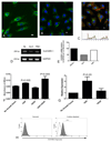
Figure 2. CLEVER-1/stabilin-1 is expressed on hepatic endothelial cells in vitro and undergoes trafficking with colocalisation to Trans Golgi Network.
(A) Representative image of immunofluorescent staining of HSEC with CLEVER-1/stabilin-1 (green) and nuclear counter stain(blue). (B) Representative image of immunofluorescent staining of HSEC after bafilomycin treatment showing CLEVER-1 localisation to intracellular vesicles (C) Confirmation of CLEVER-1/stabilin-1 in trans-Golgi network (TGN) after bafilomycin treatment; CLEVER-1/stabilin-1 (red) and TGN marker TGN-46 (green), with graphical representation below of fluorescent intensities to demonstrate areas of overlap. Bar 10µm. D) RT-PCR confirming CLEVER-1/stabilin-1 mRNA in HSEC isolated from liver tissue from normal subjects and those with liver disease (NL- normal liver, ALD-alcoholic liver disease, PBC- primary biliary cirrhosis) and (E) semiquantitative measurement of CLEVER-1 mRNA levels in these samples. (F) Cell based ELISA of HSEC stained with anti-CLEVER-1/stabilin-1 antibodies unstimulated and HGF or VEGF stimulated HSEC. Data are the mean of 3 replicate experiments and values represent the mean absorbance of three replicate wells minus the absorbance of an isotype- matched control Ab. (G) Quantitatve PCR showing relative expression of CLEVER-1 mRNA in HSEC from unstimulated HSEC and growth factor stimulated HSEC. Data are the mean of 3 independent experiments. (H) FACS analysis demonstrating surface expression CLEVER-1 on untreated and cytokine (TNFα and IFNγ) treated HSEC.
Figure 3
Figure 3. CLEVER-1/stabilin-1 mediates lymphocyte binding to hepatic sinusoids and the transmigration of lymphocytes across cytokine treated HSEC under shear flow.
(A) Static Stamper-Woodruff adhesion assays of peripheral blood lymphocyte binding to sinusoidal vessels in human liver tissue sections treated with 3-372 (anti CLEVER-1/stabilin-1) or appropriate class matched control antibody. The results of 9 independent experiments are shown as mean percentage of maximal binding +/-SEM. Bar 50µm. Blocking CLEVER-1 significantly reduced binding compared with control mAb. (B) – (D) the effect of blocking CLEVER-1/stabilin-1 on the adhesion of unfractionated peripheral blood lymphocytes to HSEC under flow are shown. (B) CLEVER-1/stabilin-1 blockade increased the number of rolling cells at low shear stress but had no effect at high shear stress. There was no alteration of cells undergoing firm adhesion or the proportion undergoing activation/shape change (C). However the proportion of adherent cells that underwent transendothelial migration through HSEC was significantly reduced by CLEVER-1/stabilin-1 blockade (C and D). The results are expressed as percentage of binding when compared with an isotype matched control. The results are mean+/- SEM of six independent experiments using different HSEC and PBLs from different donors except for experiments at high shear stress which are the mean +/- SEM of three independent experiments. The reduction in the percentage of adherent cells that transmigrated across HSEC (D) is shown for each experiment, direct comparison is made with control antibody vs CLEVER-1/stabilin-1 blockade.
Figure 4
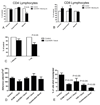
Figure 4. CLEVER-1/stabilin-1 mediates the transmigration of CD4 lymphocytes, specifically T regulatory cells, but not CD8 lymphocytes.
Flow adhesion assays using HSEC were carried out with purified peripheral blood A) CD4 and B) CD8 lymphocytes and the proportion of adherent cells, shape changed and migrated cells quantified. CLEVER-1/stabilin-1 blockade had no effect on static adhesion or shape change but significantly reduced CD4 T cells transmigration (A) but had no effect on CD8 T cells transmigration (B). Flow adhesion assays were then repeated with highly purified peripheral blood CD4 T cells sorted into either effector cells CD4+CD25- or regulatory T cells CD4+CD25+ cells. More than 90% of the CD4+CD25+ cells were also FoxP3+ and expressed low levels of CD127 consistent with a T regulatory cell phenotype. (C) The proportion of adherent T regulatory cells that transmigrated was reduced significantly by CLEVER-1/stabilin-1 blockade. The results are expressed as percentage of binding when compared with an isotype matched control. The results are mean+/- SEM of four independent experiments using different HSEC and PBLs from different donors. (D) The contribution of ICAM-1 and VAP-1 together with CLEVER-1/stabilin-1 blockade to the transendothelial migration of T regulatory cells under flow was assessed. (E) No antibody alone significantly affected adhesion but all three were implicated in transendothelial migration. The results are percentage of binding when compared with an isotype matched control shown as the mean+/- SEM of four independent experiments using different HSEC and PBLs from different donors.
Figure 5
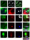
Figure 5. T regulatory cells transmigrate via the transcellular route across HSEC with enrichment of CLEVER-1/stabilin-1 during diapedesis.
Representative confocal image of CD4 lymphocytes undergoing transcellular migration. HSEC were stained with cell tracker green (A) and HSEC and lymphocyte nucleii were stained with DAPI (B). Merged image (C) demonstrates lymphocytes (arrows) clearly transmigrating through channels formed within the HSEC cytoplasm (green). Transcellular pores were enriched with a ring of ICAM-1 around transmigrating lymphocytes (D-G). VAP-1 was also detected around lymphocytes in close association with ICAM-1 (H-K). We confirmed that T regulatory cells underwent transcellular migration (L-O) with FoxP3 staining and orthogonal (XZ) projections across the plane indicated by the white line in panel O demonstrating that the T regulatory cell was crossing the endothelial cell. Staining with cell tracker green and mAbs demonstrate that CLEVER-1/stabilin-1 colocalises around FoxP3 positive cell (P-S). Orthogonal projection across the plane indicated by the white line in panel S confirms that CLEVER-1/stabilin-1 enriches around T regulatory cell during transmigration. Bar 10µm
Figure 6
Figure 6. Live cell imaging of CD4 lymphocytes undergoing transcellular migration across HSEC
To confirm that the above results were not affected by fixation artefacts we carried out live cell imaging of CD4 T cells migrating through monolayers of HSEC under flow. Imaging with confocal microscopy was able to capture lymphocytes undergoing transmigration through HSEC cytoplasm. HSEC and lymphocyte cell membranes can be seen labelled with a red dye (A, E) whilst HSEC cytoplasm was prelabelled with a green dye (B, F), lymphocytes were prelabelled with blue dye (C,G). Lymphocytes within transcellular pores were clearly seen in HSEC (D, H). Representative Z stack imaging (I) confirms lymphocytes undergoing transcellular migration. Bar 10µm

Similar articles

Cited by

References

    1. Xu XD, Ueta H, Zhou S, Shi C, Koga D, Ushiki T, Matsuno K. Trafficking of recirculating lymphocytes in the rat liver: rapid transmigration into the portal area and then to the hepatic lymph. Liver Int. 2008;28:319–330. - PubMed
    1. Lalor PF, Shields P, Grant A, Adams DH. Recruitment of lymphocytes to the human liver. Immunol Cell Biol. 2002;80:52–64. - PubMed
    1. Adams DH, Hubscher SG, Fisher NC, Williams A, Robinson M. Expression of E-selectin and E-selectin ligands in human liver inflammation. Hepatology. 1996;24:533–538. - PubMed
    1. Steinhoff G, Behrend M, Schrader B, Duijvestijn AM, Wonigeit K. Expression patterns of leukocyte adhesion ligand molecules on human liver endothelia - lack of ELAM-1 and CD62 inducibility on sinusoidal endothelia and distinct distribution of VCAM-1, ICAM-1, ICAM-2 and LFA-3. American Journal of Pathology. 1993;142:481–488. - PMC - PubMed
    1. Wong J, Johnston B, Lee SS, Bullard DC, Smith CW, Beaudet AL, Kubes P. A minimal role for selectins in the recruitment of leukocytes into the inflamed liver microvasculature. J Clin Invest. 1997;99:2782–2790. - PMC - PubMed

Publication types

MeSH terms

Substances