Farnesoid X receptor, overexpressed in pancreatic cancer with lymph node metastasis promotes cell migration and invasion
- PMID: 21364590
- PMCID: PMC3065277
- DOI: 10.1038/bjc.2011.37
Farnesoid X receptor, overexpressed in pancreatic cancer with lymph node metastasis promotes cell migration and invasion
Erratum in
- Br J Cancer. 2014 Dec 9;111(12):2381
Abstract
Background: Lymph node metastasis is one of the most important adverse prognostic factors for pancreatic cancer. The aim of this study was to identify novel lymphatic metastasis-associated markers and therapeutic targets for pancreatic cancer.
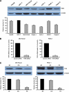
Methods: DNA microarray study was carried out to identify genes differentially expressed between 17 pancreatic cancer tissues with lymph node metastasis and 17 pancreatic cancer tissues without lymph node metastasis. The microarray results were validated by real-time PCR. Immunohistochemistry and western blotting were used to examine the expression of farnesoid X receptor (FXR). The function of FXR was studied by small interfering RNA and treatment with FXR antagonist guggulsterone and FXR agonist GW4064.
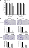
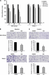
Results: Farnesoid X receptor overexpression in pancreatic cancer tissues with lymph node metastasis is associated with poor patient survival. Small interfering RNA-mediated downregulation of FXR and guggulsterone-mediated FXR inhibition resulted in a marked reduction in cell migration and invasion. In addition, downregulation of FXR reduced NF-κB activation and conditioned medium from FXR siRNA-transfected cells showed reduced VEGF levels. Moreover, GW4064-mediated FXR activation increased cell migration and invasion.
Conclusions: These findings indicated that FXR overexpression plays an important role in lymphatic metastasis of pancreatic cancer and that downregulation of FXR is an effective approach for inhibition of pancreatic tumour progression.
Figures


Similar articles
-
Inhibition of SCAMP1 suppresses cell migration and invasion in human pancreatic and gallbladder cancer cells.Tumour Biol. 2013 Oct;34(5):2731-9. doi: 10.1007/s13277-013-0825-9. Epub 2013 May 8. Tumour Biol. 2013. PMID: 23653380
-
The microRNA-218 and ROBO-1 signaling axis correlates with the lymphatic metastasis of pancreatic cancer.Oncol Rep. 2013 Aug;30(2):651-8. doi: 10.3892/or.2013.2516. Epub 2013 Jun 3. Oncol Rep. 2013. PMID: 23733161
-
Upregulated long noncoding RNA LINC01296 indicates a dismal prognosis for pancreatic ductal adenocarcinoma and promotes cell metastatic properties by affecting EMT.J Cell Biochem. 2019 Jan;120(1):552-561. doi: 10.1002/jcb.27411. Epub 2018 Sep 11. J Cell Biochem. 2019. PMID: 30203487
-
MiR-132 promotes the proliferation, invasion and migration of human pancreatic carcinoma by inhibition of the tumor suppressor gene PTEN.Prog Biophys Mol Biol. 2019 Nov;148:65-72. doi: 10.1016/j.pbiomolbio.2017.09.019. Epub 2017 Sep 20. Prog Biophys Mol Biol. 2019. PMID: 28941804 Review.
-
Molecular mechanism underlying lymphatic metastasis in pancreatic cancer.Biomed Res Int. 2014;2014:925845. doi: 10.1155/2014/925845. Epub 2014 Jan 22. Biomed Res Int. 2014. PMID: 24587996 Free PMC article. Review.
Cited by
-
Inhibition of SCAMP1 suppresses cell migration and invasion in human pancreatic and gallbladder cancer cells.Tumour Biol. 2013 Oct;34(5):2731-9. doi: 10.1007/s13277-013-0825-9. Epub 2013 May 8. Tumour Biol. 2013. PMID: 23653380
-
Guggulsterone decreases proliferation and metastatic behavior of pancreatic cancer cells by modulating JAK/STAT and Src/FAK signaling.Cancer Lett. 2013 Dec 1;341(2):166-77. doi: 10.1016/j.canlet.2013.07.037. Epub 2013 Aug 3. Cancer Lett. 2013. PMID: 23920124 Free PMC article.
-
Oxysterols and Gastrointestinal Cancers Around the Clock.Front Endocrinol (Lausanne). 2019 Jul 17;10:483. doi: 10.3389/fendo.2019.00483. eCollection 2019. Front Endocrinol (Lausanne). 2019. PMID: 31379749 Free PMC article. Review.
-
Current and future roles of mucins in cholangiocarcinoma-recent evidences for a possible interplay with bile acids.Ann Transl Med. 2018 Sep;6(17):333. doi: 10.21037/atm.2018.07.16. Ann Transl Med. 2018. PMID: 30306072 Free PMC article. Review.
-
Expression of NR1H3 in endometrial carcinoma and its effect on the proliferation of Ishikawa cells in vitro.Onco Targets Ther. 2019 Jan 18;12:685-697. doi: 10.2147/OTT.S180534. eCollection 2019. Onco Targets Ther. 2019. PMID: 30705597 Free PMC article.
References
-
- Aghdassi A, Phillips P, Dudeja V, Dhaulakhandi D, Sharif R, Dawra R, Lerch MM, Saluja A (2007) Heat shock protein 70 increases tumorigenicity and inhibits apoptosis in pancreatic adenocarcinoma. Cancer Res 67: 616–625 - PubMed
-
- Arinaga M, Noguchi T, Takeno S, Chujo M, Miura T, Uchida Y (2003) Clinical significance of vascular endothelial growth factor C and vascular endothelial growth factor receptor 3 in patients with nonsmall cell lung carcinoma. Cancer 97: 457–464 - PubMed
-
- De Gottardi A, Dumonceau JM, Bruttin F, Vonlaufen A, Morard I, Spahr L, Rubbia-Brandt L, Frossard JL, Dinjens WN, Rabinovitch PS, Hadengue A (2006) Expression of the bile acid receptor FXR in Barrett's esophagus and enhancement of apoptosis by guggulsterone in vitro. Mol Cancer 5: 48. - PMC - PubMed
-
- De Gottardi A, Touri F, Maurer CA, Perez A, Maurhofer O, Ventre G, Bentzen CL, Niesor EJ, Dufour JF (2004) The bile acid nuclear receptor FXR and the bile acid binding protein IBABP are differently expressed in colon cancer. Dig Dis Sci 49: 982–989 - PubMed
Publication types
MeSH terms
Substances
Grants and funding
LinkOut - more resources
Full Text Sources
Medical

