. 2011 Feb 10;7(2):e1001278.
doi: 10.1371/journal.ppat.1001278.

NS2 protein of hepatitis C virus interacts with structural and non-structural proteins towards virus assembly

Affiliations

NS2 protein of hepatitis C virus interacts with structural and non-structural proteins towards virus assembly

Costin-Ioan Popescu et al. PLoS Pathog. .

Abstract

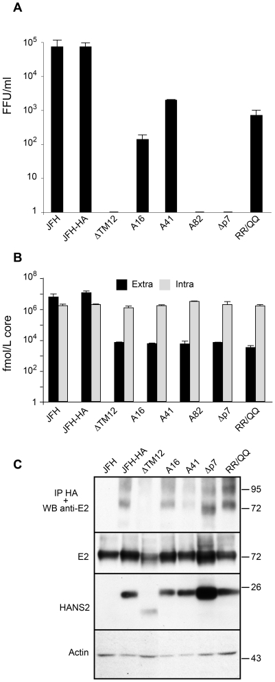
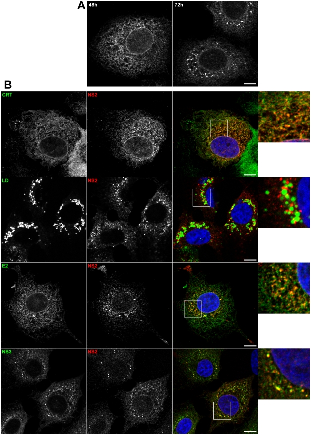
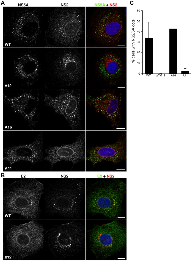
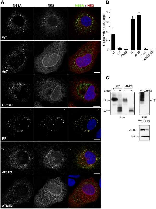
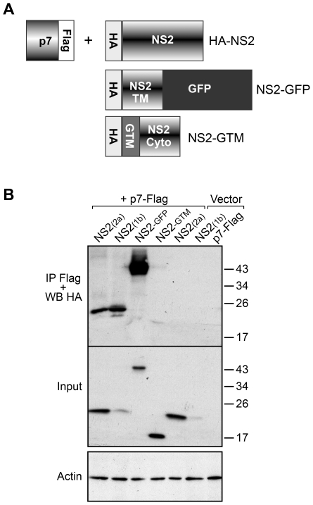
Growing experimental evidence indicates that, in addition to the physical virion components, the non-structural proteins of hepatitis C virus (HCV) are intimately involved in orchestrating morphogenesis. Since it is dispensable for HCV RNA replication, the non-structural viral protein NS2 is suggested to play a central role in HCV particle assembly. However, despite genetic evidences, we have almost no understanding about NS2 protein-protein interactions and their role in the production of infectious particles. Here, we used co-immunoprecipitation and/or fluorescence resonance energy transfer with fluorescence lifetime imaging microscopy analyses to study the interactions between NS2 and the viroporin p7 and the HCV glycoprotein E2. In addition, we used alanine scanning insertion mutagenesis as well as other mutations in the context of an infectious virus to investigate the functional role of NS2 in HCV assembly. Finally, the subcellular localization of NS2 and several mutants was analyzed by confocal microscopy. Our data demonstrate molecular interactions between NS2 and p7 and E2. Furthermore, we show that, in the context of an infectious virus, NS2 accumulates over time in endoplasmic reticulum-derived dotted structures and colocalizes with both the envelope glycoproteins and components of the replication complex in close proximity to the HCV core protein and lipid droplets, a location that has been shown to be essential for virus assembly. We show that NS2 transmembrane region is crucial for both E2 interaction and subcellular localization. Moreover, specific mutations in core, envelope proteins, p7 and NS5A reported to abolish viral assembly changed the subcellular localization of NS2 protein. Together, these observations indicate that NS2 protein attracts the envelope proteins at the assembly site and it crosstalks with non-structural proteins for virus assembly.


Conflict of interest statement

The authors have declared that no competing interests exist.

Figures

Figure 1
Figure 1. Rationale for mutagenesis in NS2 transmembrane region.
(A) Schematic representation of the topology of E2, p7 and NS2 proteins. (B) Position of the inserted alanine residues in the putative N-terminal membrane domain of NS2. Alignments of NS2 membrane domain sequences from HCV strains JFH1 (genotype 2a, accession number AB047639) and Con1 (genotype 1b, AJ238799). Amino acids are numbered with respect to NS2 and the HCV JFH1 polyprotein (top row). Second. struct., secondary structure deduced from the NMR analyses of NS2 synthetic peptides from Con1 strain (Jirasko et.al. the 16th international Conference on HCV and Related Viruses, Nice, October 3–7, 2009); c = coil, h = helix; capital letters indicate canonical helix structure. Predicted TM, consensus transmembrane (TM) segment predictions were deduced from a set of 6 available web-based algorithms prediction methods (DAS, TOPPRED2, TMHMM, SOSUI, TMPRED, PHD-TM) and represented by stretches of “T”. Arrows indicate the positions of the various alanine insertions. (C) Ribbon representations of the molecular homology model of NS2[1–27] of JFH1 (left) and the theoretical model for alanine insertion mutant A16 (right). An Ala insertion (shown in magenta) twists the helix by 110°. The N-terminal part of the model is shown in the same orientation as in the left model to highlight the distortion of residue positions on the C-terminal part of the helix. The side chains of indicated residues are shown to highlight this distortion. These models were constructed by using the NMR structure of Con1 NS2[1–27] (; PDB entry, 2JY0) as template and the Swiss-PdbViewer program (http://www.expasy.ch/swissmod/). Residues are colored based on the chemical properties of their side chains: hydrophobic (gray) and polar (yellow). Acidic (Asp) and basic (Arg, Lys) residues are red and blue, respectively. His is cyan, and Gly is light gray. The membrane interfaces and hydrophobic core are schematically represented.
Figure 2
Figure 2. NS2 transmembrane region enables the NS2-E2 interaction.
(A) Mutations in NS2 transmembrane region drastically decrease the production of infectious virions. Huh-7 cells were electroporated with viral RNA transcribed from different JFH-1 derived mutants. At 72h post-electroporation, the virus infectivity present in the supernatants was determined by titration of foci forming units (FFUs). Error bars indicate SD from at least two independent experiments performed in duplicate. A16, A41 and A82 correspond to JFH-HA virus with an alanine residue inserted at position 16, 41 and 82 of NS2, respectively. The following viruses were also analyzed in parallel: JFH-ΔTM12-HA (ΔTM12), JFH-Δp7-HA (Δp7) and JFH-RR/QQ-HA (RR/QQ). (B) The NS2 mutations affect the viral secretion, but not the replication capacity. Huh-7 cells were electroporated with viral RNA transcribed from different JFH-1 derived mutants. At 72h post-electroporation, the amount of extracellular and intracellular core antigen was determined in supernatants and cell lysates, respectively. Error bars indicate SD from at least two independent experiments performed in duplicate. (C) HA-NS2 co-immunoprecipitates with E2. Huh-7 cells were electroporated with viral RNA transcribed from different JFH-1 derived mutants. At 72h post-electroporation, cells were lysed and immunoprecipitation was performed with an anti-HA antibody. The immunoprecipitates were separated by SDS-PAGE and analyzed by Western blotting with an anti-E2 antibody. The presence of E2, and HA-NS2 in electroporated cells was confirmed by Western blotting and the actin content was also analyzed to verify that equal amounts of cell lysates have been loaded. It has to be noted that the E2 band corresponds to both E2 and E2p7 proteins. However, in the absence of p7, the E2-p7 form is absent.
Figure 3
Figure 3. Subcellular localization of NS2.
(A) NS2 accumulates in dotted structures. Huh-7 cells electroporated with JFH-HA RNA were grown on coverslips and fixed at 48h and 72h post-electroporation. The subcellular localization of HA-NS2 was analyzed by immunofluorescence using an anti-HA antibody. (B) Colocalization of NS2 dotted structures with cellular and viral markers. JFH-HA electroporated cells grown on coverslips were fixed at 72h post-electroporation and processed for double-label immunofluorescence for HA-NS2 (red) and the ER marker calreticulin (CRT in green) or the LDs stained with BODIPY 493/503 (green). JFH-HA electroporated cells were further stained for HA-NS2 (red) and HCV proteins NS3 or E2 (green). The nuclei were stained with DAPI. Representative confocal images of individual cells are shown in grey and the merge images in color. Zoomed views of the indicated areas are shown in the right column. Bar, 10 µm.
Figure 4
Figure 4. NS2 colocalization with viral proteins in association with LDs.
JFH-HA electroporated cells grown on coverslips were fixed at 72h post-electroporation and processed for triple-label immunofluorescence for HA-NS2 (red), the LDs (green), and (A) NS5A or (B) core protein (C) (blue). Individual confocal images of each labeling are shown in left panels, with a merged image of the three channels in the top left panel. Two by two overlays are shown in right panels. A view of the entire cell is shown in the top right panel. Bar, 10 µm. (C) The kinetics of virus production for JFH-HA. Huh-7 cells were electroporated with viral RNA transcribed from JFH-HA construct. At different time points post-electroporation, the virus infectivity present in the supernatants was determined by titration of foci forming units (FFUs)(upper panel). Error bars indicate SE from at least two independent experiments performed in duplicate. The kinetics of NS2/NS5A positive dots parallels virus production (lower panel). Huh-7 cells electroporated with JFH-HA RNA were grown on coverslips and fixed at different time points post-electroporation and labeled with anti-HA and anti-NS5A antibodies. The cells which presented at least 3 dots NS2/NS5A positive were considered positive for NS2 dotted structures. The results were expressed as percentage of total counted infected cells. At least 300 infected cells were counted for each time point. Error bars indicate SE from at least two independent experiments. (D) NS2 detection by immuno-EM microscopy. JFH-HA electroporated cells grown in 75 cm2 flasks were fixed at 72h post-electroporation and processed for immuno-electron microscopy labeling with anti-HA antibody. A representative image is shown where three HA-NS2 clusters (arrows) could be observed in the proximity of a LD. Two of these clusters of gold particles (white arrows) are lying on well-preserved ER bilayers. For one of these clusters of gold particles, a connection between the ER bilayer and the LD could be observed (white arrowhead). A third cluster of gold particles (black arrow) is observed on a less preserved ER bilayer.
Figure 5
Figure 5. Subcellular localization of NS2 mutants in TM region.
(A) and (B) Effect of mutations on the accumulation of NS2 in dotted structures. Huh-7 cells electroporated with JFH-HA RNA (WT) or mutant genomes were grown on coverslips and fixed at 72h post-electroporation. The following viruses were analyzed: JFH-HA (WT), JFH-?TM12-HA (?TM12), JFH-A16-HA (A16) and JFH-A41-HA (A41). The subcellular localization of HA-NS2 was analyzed by immunofluorescence using anti-HA (red), anti-NS5A (green) (Panel A) and anti-E2 (green) (Panel B) antibodies. The nuclei were stained with DAPI. Representative confocal images of NS2 and NS5A labelings are shown in grey, and the merge images in color. Bar, 10 micro µm. (C) Mutations in NS2 transmembrane region drastically decrease the number of cells presenting NS2 dotted structures. Huh-7 cells electroporated with JFH-HA RNA were grown on coverslips and fixed at different time points post-electroporation and labeled with anti-HA and anti-NS5A antibodies. Cells showing at least 3 NS2/NS5A dots were considered positive for NS2 dotted structures. The results were expressed as percentage of total counted cells. At least 250 infected cells were counted. Error bars indicate SD from at least two independent experiments.
Figure 6
Figure 6. Subcellular localization of NS2 in assembly deficient mutants in the structural region.
(A) Effect of mutations on the accumulation of NS2 in dotted structures. Huh-7 cells electroporated with wild-type or mutant genomes were grown on coverslips and fixed at 72h post-electroporation. The subcellular localization of HA-NS2 was analyzed by immunofluorescence using anti-HA (red) and anti-NS5A (green) antibodies. The nuclei were stained with DAPI. The following viruses were analyzed: JFH-HA (WT), JFH-Δp7-HA (Δp7), JFH-RR/QQ-HA (RR/QQ), JFH-HA-PP (PP), JFH-ΔE1E2-HA (ΔE1E2) and JFH-ΔTME2-HA (ΔTME2). Representative confocal images of NS2 and NS5A labelings are shown in grey, and the merge images in color. Bar, 10 µm. (B) Mutations in the structural region of HCV have different effects upon the number of cells presenting NS2 dotted structures. Huh-7 cells electroporated with JFH-HA RNA were grown on coverslips and fixed at different time points post-electroporation and labeled with anti-HA and anti-NS5A antibodies. Cells showing at least 3 NS2/NS5A dots were considered positive for NS2 dotted structures. The results were expressed as percentage of total counted cells. At least 220 infected cells were counted. Error bars indicate SD from at least two independent experiments. In this experiment, JFH-ΔE1E2TME2-HA (ΔE1E2TME2) construct was used in addition to the above mentioned viruses. (C) The transmembrane domain of E2 mediates the NS2-E2 interaction. Huh-7 cells were electroporated with viral RNA transcribed from JFH-HA or JFH-ΔTME2-HA (ΔTME2) mutant. At 72h post-electroporation, cells were lysed and immunoprecipitation was performed with an anti-HA antibody. The immunoprecipitates were separated by SDS-PAGE and analyzed by Western blotting with an anti-E2 antibody. Glycosylated or deglycosylated lysates were blotted against E2 and HA-NS2. The actin content was also analyzed to verify that equal amounts of cell lysates have been loaded.
Figure 7
Figure 7. NS2 and p7 interact in a co-immunoprecipitation assay.
(A) Schematic representation of the constructs used in this study. NS2-GFP corresponds to the transmembrane domain of NS2 in fusion with GFP, whereas NS2-GTM corresponds to the transmembrane domain of VSV-G protein in fusion with the cytosolic domain of NS2. All the proteins contain a HA tag at their N-terminus. (B) HA-NS2 co-immunoprecipitates with p7-Flag. 293T cells were transfected with plasmids expressing p7-Flag, HA-NS2 from different genotypes, HA-NS2 mutants or control plasmids. At 24h post-transfection cells were lysed and immunoprecipitations with an anti-Flag antibody were performed. The immunoprecipitates were separated by SDS-PAGE and analyzed by Western blotting with an anti-HA antibody to identify the presence of co-immunoprecipitated HA-NS2. The presence of HA-NS2 in transfected cells was confirmed by Western blotting. The actin content was also analyzed to verify that equal amounts of cell lysates have been used.
Figure 8
Figure 8. NS2 and p7 interact in FRET-FLIM analyses.
(A) Schematic representation of the constructs used in this study. (B) Immunofluorescence analysis of the co-expression of CFP-p7 and YFP-NS2. U2OS cells were co-transfected with plasmids expressing CFP-p7 and YFP-NS2. At 24h post-transfection, the subcellular localization of the different proteins was assessed by confocal microscopy. (C) Western blot analysis of the expression of CFP-p7, YFP-NS2 and CFP-EYF. U2OS cells were transfected with plasmids expressing CFP-p7, YFP-NS2 or CFP-EYF. At 24h post-transfection, cells were lysed and protein expression was confirmed by SDS-PAGE followed by Western blotting. (D) FLIM analyses. Samples were subjected to FLIM and color coded maps were obtained. The regions where the FRET events are present are marked with squares. The colors represent the progression from minimum (yellow) to maximum (blue) fluorescence lifetime.
Figure 9
Figure 9. Subcellular localization of NS2 in assembly deficient mutants in NS5A protein.
(A, B) Phenotype of the NS5A mutants. Virus infectivity (A), extra (black bars) and intracellular (light grey bars) core determination (B) were performed as described in Figure 2. The following viruses were analyzed: JFH-HA (WT), JFH-S/A-HA (S/A), JFH-3BS/A-HA (3BS/A) and JFH-S/D-HA (S/D). In addition, JFH-HA-PP (PP) was also analyzed in parallel. (C) Analysis of the expression and phosphorylation of the NS5A mutants. The hyperphosphorylated form of NS5A is indicated by an asterisk. The presence of core and NS5A was confirmed by Western blotting and the actin content was also analyzed to verify that equal amounts of cell lysates have been loaded. (D) Effect of mutations on the accumulation of NS2 in dotted structures. Huh-7 cells electroporated with JFH-HA RNA (WT) or mutant genomes were grown on coverslips and fixed at 72h post-electroporation. The subcellular localization of HA-NS2 was analyzed by immunofluorescence using anti-HA (red) and anti-NS5A (green) antibodies. The nuclei were stained with DAPI. Representative confocal images of NS2 and NS5A labelings are shown in grey, and the merge images in color. Bar, 10 µm. (E) Mutations in NS5A protein drastically decrease the number of cells presenting NS2 dotted structures. Huh-7 cells electroporated with JFH-HA RNA or the indicated mutants were grown on coverslips, fixed at 72 h post-electroporation and labeled with anti-HA and anti-NS5A antibodies. Cells showing at least 3 dots NS2/NS5A positive were considered positive for NS2 dotted structures. The results were expressed as percentage of total counted cells (at least 228). Error bars indicate SD from at least two independent experiments.
Figure 10
Figure 10. Model of NS2 role in the assembly process.
Upon viral polyprotein translation and processing, three viral modules are formed: the core protein (C), the replication complex (RC) and the E1E2p7NS2 complex. E1E2p7NS2 complex assembles through the interaction of E1E2 heterodimer and p7NS2 unit (4) and migrates close to the RC independently of core protein due to signals present in p7NS2 and E2 (3). The core protein localizes around the LDs (1) where it recruits the RC by core-NS5A interaction (2).

Similar articles

Cited by

References

    1. Wasley A, Alter MJ. Epidemiology of hepatitis C: geographic differences and temporal trends. Semin Liver Dis. 2000;20:1–16. - PubMed
    1. Feld JJ, Hoofnagle JH. Mechanism of action of interferon and ribavirin in treatment of hepatitis C. Nature. 2005;436:967–972. - PubMed
    1. Lindenbach BD, Thiel HJ, Rice CM. Flaviviridae: the viruses and their replication. In: Knipe DM, Howley PM, editors. Fields Virology. 5th ed. Philadelphia, Pa: Lippincott Williams & Wilkins; 2007. pp. 1101–1152.
    1. Moradpour D, Penin F, Rice CM. Replication of hepatitis C virus. Nat Rev Microbiol. 2007;5:453–463. - PubMed
    1. Blight KJ, McKeating JA, Rice CM. Highly permissive cell lines for subgenomic and genomic hepatitis C virus RNA replication. J Virol. 2002;76:13001–13014. - PMC - PubMed

Publication types

MeSH terms

Substances

LinkOut - more resources