The ability of pandemic influenza virus hemagglutinins to induce lower respiratory pathology is associated with decreased surfactant protein D binding
- PMID: 21334038
- PMCID: PMC3060949
- DOI: 10.1016/j.virol.2011.01.029
The ability of pandemic influenza virus hemagglutinins to induce lower respiratory pathology is associated with decreased surfactant protein D binding
Abstract
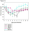
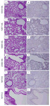
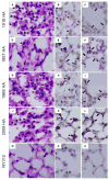
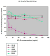
Pandemic influenza viral infections have been associated with viral pneumonia. Chimeric influenza viruses with the hemagglutinin segment of the 1918, 1957, 1968, or 2009 pandemic influenza viruses in the context of a seasonal H1N1 influenza genome were constructed to analyze the role of hemagglutinin (HA) in pathogenesis and cell tropism in a mouse model. We also explored whether there was an association between the ability of lung surfactant protein D (SP-D) to bind to the HA and the ability of the corresponding chimeric virus to infect bronchiolar and alveolar epithelial cells of the lower respiratory tract. Viruses expressing the hemagglutinin of pandemic viruses were associated with significant pathology in the lower respiratory tract, including acute inflammation, and showed low binding activity for SP-D. In contrast, the virus expressing the HA of a seasonal influenza strain induced only mild disease with little lung pathology in infected mice and exhibited strong in vitro binding to SP-D.
Published by Elsevier Inc.
Figures


Similar articles
-
Influenza Virus Hemagglutinins H2, H5, H6, and H11 Are Not Targets of Pulmonary Surfactant Protein D: N-Glycan Subtypes in Host-Pathogen Interactions.J Virol. 2020 Feb 14;94(5):e01951-19. doi: 10.1128/JVI.01951-19. Print 2020 Feb 14. J Virol. 2020. PMID: 31826991 Free PMC article.
-
Contemporary avian influenza A virus subtype H1, H6, H7, H10, and H15 hemagglutinin genes encode a mammalian virulence factor similar to the 1918 pandemic virus H1 hemagglutinin.mBio. 2014 Nov 18;5(6):e02116. doi: 10.1128/mBio.02116-14. mBio. 2014. PMID: 25406382 Free PMC article.
-
N-linked glycosylation of the hemagglutinin protein influences virulence and antigenicity of the 1918 pandemic and seasonal H1N1 influenza A viruses.J Virol. 2013 Aug;87(15):8756-66. doi: 10.1128/JVI.00593-13. Epub 2013 Jun 5. J Virol. 2013. PMID: 23740978 Free PMC article.
-
The 1918 Influenza Virus PB2 Protein Enhances Virulence through the Disruption of Inflammatory and Wnt-Mediated Signaling in Mice.J Virol. 2015 Dec 9;90(5):2240-53. doi: 10.1128/JVI.02974-15. J Virol. 2015. PMID: 26656717 Free PMC article.
-
The number and position of N-linked glycosylation sites in the hemagglutinin determine differential recognition of seasonal and 2009 pandemic H1N1 influenza virus by porcine surfactant protein D.Virus Res. 2012 Oct;169(1):301-5. doi: 10.1016/j.virusres.2012.08.003. Epub 2012 Aug 15. Virus Res. 2012. PMID: 22921759
Cited by
-
An Ultrasensitive Mechanism Regulates Influenza Virus-Induced Inflammation.PLoS Pathog. 2015 Jun 5;11(6):e1004856. doi: 10.1371/journal.ppat.1004856. eCollection 2015 Jun. PLoS Pathog. 2015. PMID: 26046528 Free PMC article.
-
Pathogenicity and transmissibility of North American triple reassortant swine influenza A viruses in ferrets.PLoS Pathog. 2012;8(7):e1002791. doi: 10.1371/journal.ppat.1002791. Epub 2012 Jul 19. PLoS Pathog. 2012. PMID: 22829764 Free PMC article.
-
A Role for Neutrophils in Viral Respiratory Disease.Front Immunol. 2017 May 12;8:550. doi: 10.3389/fimmu.2017.00550. eCollection 2017. Front Immunol. 2017. PMID: 28553293 Free PMC article. Review.
-
The Role and Molecular Mechanism of Action of Surfactant Protein D in Innate Host Defense Against Influenza A Virus.Front Immunol. 2018 Jun 13;9:1368. doi: 10.3389/fimmu.2018.01368. eCollection 2018. Front Immunol. 2018. PMID: 29951070 Free PMC article. Review.
-
Neuraminidase Activity and Resistance of 2009 Pandemic H1N1 Influenza Virus to Antiviral Activity in Bronchoalveolar Fluid.J Virol. 2016 Apr 14;90(9):4637-4646. doi: 10.1128/JVI.00013-16. Print 2016 May. J Virol. 2016. PMID: 26912622 Free PMC article.
References
-
- Brown-Augsburger P, Chang D, Rust K, Crouch EC. Biosynthesis of surfactant protein D. Contributions of conserved NH2-terminal cysteine residues and collagen helix formation to assembly and secretion. J Biol Chem. 1996;271(31):18912–9. - PubMed
-
- Bush RM, Bender CA, Subbarao K, Cox NJ, Fitch WM. Predicting the evolution of human influenza A. Science. 1999;286(5446):1921–5. - PubMed
-
- CDC . CDC Estimates of 2009 H1N1 Influenza Cases, Hospitalizations and Deaths in the United States, April 2009 – February 13, 2010. Vol. 2010. CDC; Atlanta: 2010.
-
- Cottey R, Rowe CA, Bender BS. Influenza Virus. In: Coico R, editor. Current Protocols in Immunology. John Wiley and Sons; Hoboken, NJ: 2003. pp. 19.11.6–19.11.9.pp. 19.11.19–19.11.20. - PubMed
Publication types
MeSH terms
Substances
Grants and funding
LinkOut - more resources
Full Text Sources

