. 2011 May;85(9):4122-34.
doi: 10.1128/JVI.02232-10. Epub 2011 Feb 16.

Evidence that TMPRSS2 activates the severe acute respiratory syndrome coronavirus spike protein for membrane fusion and reduces viral control by the humoral immune response

Affiliations

Evidence that TMPRSS2 activates the severe acute respiratory syndrome coronavirus spike protein for membrane fusion and reduces viral control by the humoral immune response

Ilona Glowacka et al. J Virol. 2011 May.

Abstract

The spike (S) protein of the severe acute respiratory syndrome coronavirus (SARS-CoV) can be proteolytically activated by cathepsins B and L upon viral uptake into target cell endosomes. In contrast, it is largely unknown whether host cell proteases located in the secretory pathway of infected cells and/or on the surface of target cells can cleave SARS S. We along with others could previously show that the type II transmembrane protease TMPRSS2 activates the influenza virus hemagglutinin and the human metapneumovirus F protein by cleavage. Here, we assessed whether SARS S is proteolytically processed by TMPRSS2. Western blot analysis revealed that SARS S was cleaved into several fragments upon coexpression of TMPRSS2 (cis-cleavage) and upon contact between SARS S-expressing cells and TMPRSS2-positive cells (trans-cleavage). cis-cleavage resulted in release of SARS S fragments into the cellular supernatant and in inhibition of antibody-mediated neutralization, most likely because SARS S fragments function as antibody decoys. trans-cleavage activated SARS S on effector cells for fusion with target cells and allowed efficient SARS S-driven viral entry into targets treated with a lysosomotropic agent or a cathepsin inhibitor. Finally, ACE2, the cellular receptor for SARS-CoV, and TMPRSS2 were found to be coexpressed by type II pneumocytes, which represent important viral target cells, suggesting that SARS S is cleaved by TMPRSS2 in the lung of SARS-CoV-infected individuals. In summary, we show that TMPRSS2 might promote viral spread and pathogenesis by diminishing viral recognition by neutralizing antibodies and by activating SARS S for cell-cell and virus-cell fusion.


Figures
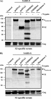
Fig. 1.
Fig. 1.
Proteolytic processing of SARS S by TMPRSS2. (Top) VLPs were produced by coexpression of HIV p55 Gag and SARS S in the absence and presence of coexpressed human TMPRSS2, TMPRSS4, or murine matriptase-3, treated with trypsin or PBS, and analyzed for S protein and HIV p55 Gag content as indicated, using a serum specific for the S2 subunit of SARS S. (B) The experiment was carried out as described for panel A. However, an S1-specific antiserum was used for SARS S protein detection.
Fig. 2.
Fig. 2.
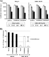
Impact of SARS S processing by TMPRSS2 on cathepsin dependence and neutralization sensitivity. (A) 293T cells engineered to express large amounts of ACE2 were incubated with the indicated concentrations of the cathepsin B/L inhibitor MDL 28170 and inoculated with pseudotypes in triplicate, and luciferase activities were determined at 72 h postinfection. Activities measured in the absence of inhibitor were set as 100%. A representative experiment out of three is shown; error bars indicate standard deviations. In the absence of inhibitor, the following luciferase counts were measured: VSV-G, 55,642 ± 4877 cps; SARS S plus pcDNA3, 64,751 ± 11,505 cps; SARS S plus TMPRSS2, 59,071 ± 5,087 cps; SARS plus TMPRSS4, 94,684 ± 4,576 cps. (B) Equal volumes of pseudotypes bearing SARS S wt were incubated for 60 min with the indicated dilutions of the sera R1 and R1LI1 in triplicate and then added to ACE2-expressing 293T cells. Luciferase activities in cell lysates were determined after 72 h, and activities measured in the absence of serum were set as 100%. The results ± standard deviations of a representative experiment are shown. Similar results were obtained in two independent experiments. In the absence of serum, the following luciferase counts were measured: VSV-G, 9,037,372 ± 33,0551 cps; SARS-S plus pcDNA3, 5,411,448 ± 304,990 cps; SARS-S plus TMPRSS2, 444,923 ± 27,314 cps.
Fig. 3.
Fig. 3.
Cleavage by TMPRSS2 induces SARS S shedding. (A) VLPs were produced in 293T cells in the absence and presence of TMPRSS2, concentrated by ultrafiltration, treated with PNGase F to remove N-linked glycans, and analyzed for SARS S and Gag protein content by Western blotting. Results of a single gel are shown, from which irrelevant lanes were removed. (B) The experiment was carried out as in panel A, but VLPs were additionally concentrated by ultracentrifugation through a 20% sucrose cushion. (C) VLPs were produced as described in panel A and then subjected to ultrafiltration followed by ultracentrifugation. Western blot analysis was employed to determine the effect of these procedures on the concentrations of SARS S and Gag protein in the VLP preparations. Ultrafiltration, VLP preparation subjected to ultrafiltration; UC pellet, VLP preparation subjected to ultrafiltration followed by ultracentrifugation and Western blot analysis of the pellets; UC supernatant, VLP preparation subjected to ultrafiltration followed by ultracentrifugation and Western blot analysis of the supernatants of ultracentrifuge reactions.
Fig. 4.
Fig. 4.
Shedding of SARS S by TMPRSS2 confers neutralization resistance. (A) The pseudoparticles indicated were produced in 293T cells in the presence or absence of TMPRSS2, concentrated by ultracentrifugation through a 20% sucrose cushion, preincubated with the indicated dilutions of the sera R1 and R1LI1, and then used for triplicate infections of 293T-ACE2 cells. Luciferase activities in cell lysates were determined after 72 h, and activities measured in the absence of serum were set as 100%. The results of a representative experiment are shown; error bars indicate standard deviations. The results were confirmed in two independent experiments. In the absence of serum, the following luciferase counts were measured: VSV-G, 509,961 ± 37,823 cps; SARS S plus pcDNA3, 497,873 ± 52,794 cps; SARS S plus TMPRSS2, 608,600 ± 97,835 cps. (B) The concentrated pseudotypes described in panel A were analyzed by Western blotting for the presence of SARS S (employing an S1-specific rabbit serum). (C) 293T cells were transiently cotransfected with expression plasmids for SARS S and TMPRSS2 or cotransfected with SARS S plasmid and empty vector (pcDNA3), and culture supernatants were harvested at 48 h after transfection. Subsequently, the supernatants were concentrated by ultrafiltration followed by ultracentrifugation through a 20% sucrose cushion. The presence of soluble SARS S protein in the supernatants of ultracentrifuged samples was analyzed by immunoblotting, as described in panel A. (D) Pseudoparticles bearing SARS S and purified by ultracentrifugation through a sucrose cushion were preincubated with the indicated dilutions of supernatants described in panel C and a 1:50 dilution of the sera R1 and R1LI1 for 60 min before addition to target cells (pcDNA3, supernatants from cells cotransfected with SARS-S and empty plasmid; TMPRSS2, supernatants from cells cotransfected with TMPRSS2 and SARS S expression plasmids). Luciferase activities in cell lysates were determined after 72 h. The results ± standard deviations of a representative experiment carried out in triplicate are shown; activities measured in the absence of serum, 1,307,409 ± 328118 cps, were set as 100%. The results were confirmed in two separate experiments.
Fig. 5.
Fig. 5.
TMPRSS2 cleaves SARS S in trans and activates SARS S for cell-cell fusion. (A) Effector cells were transfected with SARS S expression plasmid and mixed with target cells transfected with empty plasmid (pcDNA) or plasmids encoding TMPRSS2 or TMPRSS4. Lysates and supernatants of these cell mixtures were analyzed for SARS S cleavage by Western blotting, employing sera directed against the S1 and the S2 portions of SARS S for detection. Before analysis by Western blotting, supernatants were concentrated by ultrafiltration and ultracentrifugation. Cell pellet, lysates of transfected cells analyzed by Western blotting; Ultrafiltration, culture supernatants subjected to ultrafiltration followed by Western blot analysis; UC pellet, culture supernatants subjected to ultrafiltration followed by ultracentrifugation and Western blot analysis of the pellets; UC supernatant, culture supernatants subjected to ultrafiltration followed by ultracentrifugation and Western blot analysis of the supernatants of ultracentrifuge reactions. (B) Effector cells cotransfected with pGAL4-VP16 expression plasmid and either empty plasmid or SARS S expression plasmid were mixed with target cells cotransfected with the indicated plasmids and a plasmid encoding luciferase under the control of a promoter with multiple GAL4 binding sites. The cell mixtures were then treated with either PBS or trypsin, and the luciferase activities in cell lysates were quantified at 48 h after cell mixing. The results of a representative experiment performed in triplicates are shown; error bars indicate standard deviations. Similar results were observed in two independent experiments. (C) Vero E6 cells transfected with TMPRSS2 or TMPRSS4 expression plasmid or empty vector (pcDNA) were infected with SARS-CoV (Frankfurt strain 1) at an MOI of 0.1. At 29 h postinfection the cells were fixed and analyzed by microscopy. Bar, 20 μm. Arrows indicate syncytia. Similar results were obtained in an independent experiment.
Fig. 6.
Fig. 6.
Expression of TMPRSS2 on target cells reduces the requirement for acidic pH and cathepsin activity for SARS S-driven infectious entry. (A) The indicated proteases were expressed in 293T-hACE2 cells, and the cells were pretreated with medium containing the indicated concentrations of NH4Cl and MDL 28170. Subsequently, the cells were infected with pseudotypes bearing SARS S in the presence of inhibitor. The infection medium was replaced by fresh medium without inhibitor at 16 h postinfection, and the luciferase activities in cell lysates were analyzed at 72 h postinfection. The results of representative experiments performed in triplicates are shown; error bars indicate standard deviations. Similar results were obtained in two independent experiments. (B) The indicated proteases were expressed in 293T-hACE2 cells, and the cells were treated with the cathepsin inhibitor MDL 28170 before infection with SARS-CoV (Frankfurt strain 1) at an MOI of 0.1. At 5 h postinfection the cells were washed with PBS and lysed, and total RNA was extracted. SARS-CoV entry was analyzed by real-time RT-PCR specific for the N gene mRNA. Average CT values of a single experiment performed in triplicates were normalized by subtracting the respective CT values for TATA-box binding protein (reference gene). For clarity, values were subtracted by a fixed number (20). A CT difference of 3 correlated approximately with a 10-fold increase in transcripts as determined by a dilution series of both targets. Similar results were obtained in an independent experiment.
Fig. 7.
Fig. 7.
Coexpression of ACE2 and TMPRSS2 on type II pneumocytes. (A) The amount of Tmprss2 transcript in the indicated organs was quantified by PCR, employing GUSB as a housekeeping reference. The averages of three independent experiments are shown; error bars indicate standard deviations (std err). No specific signal was measured for brain in three out of three experiments. A specific signal was measured for heart in one out of three independent experiments. (B, i) Hematoxylin-and-eosin-stained section of normal lung showing several alveolar spaces, in which alveolar macrophages (M), type I pneumocytes (P1), and type II pneumocytes (P2) are labeled. Scale bar, 20 μm. (ii) Serial section of frame i immunostained for TMPRSS2 using the peroxidase technique (brown) shows strong positive staining in type II pneumocytes and alveolar macrophages. (iii) Serial section of frame ii immunostained for ACE-2 shows strong positive staining in type II pneumocytes and alveolar macrophages. (iv) Serial section of frame iii immunostained with an irrelevant mouse primary antibody (melan-A), as a negative control for frame ii, shows no immunostaining. Alveolar macrophages show a faint brown tint, due to the presence of carbon, but not the strong brown staining of macrophages seen in frame ii. (v) Serial section of frame iv immunostained using goat polyclonal serum as a primary antibody, as a negative control for frame iii. Alveolar macrophages show a faint brown tint, due to the presence of carbon, but not the strong brown staining of macrophages seen in frames ii and iii.

Similar articles

Cited by

References

    1. Babcock G. J., Esshaki D. J., Thomas W. D., Jr., Ambrosino D. M. 2004. Amino acids 270 to 510 of the severe acute respiratory syndrome coronavirus spike protein are required for interaction with receptor. J. Virol. 78:4552–4560 - PMC - PubMed
    1. Belouzard S., Chu V. C., Whittaker G. R. 2009. Activation of the SARS coronavirus spike protein via sequential proteolytic cleavage at two distinct sites. Proc. Natl. Acad. Sci. U. S. A. 106:5871–5876 - PMC - PubMed
    1. Bergeron E., et al. 2005. Implication of proprotein convertases in the processing and spread of severe acute respiratory syndrome coronavirus. Biochem. Biophys. Res. Commun. 326:554–563 - PMC - PubMed
    1. Bertram S., et al. 2010. TMPRSS2 and TMPRSS4 facilitate trypsin-independent spread of influenza virus in Caco-2 cells. J. Virol. 84:10016–10025 - PMC - PubMed
    1. Bertram S., Glowacka I., Steffen I., Kuhl A., Pöhlmann S. 2010. Novel insights into proteolytic cleavage of influenza virus hemagglutinin. Rev. Med. Virol. 20:298–310 - PMC - PubMed

Publication types

MeSH terms

Substances

LinkOut - more resources