

Preliminary Characterization of the Transcriptional Response of the Porcine Intestinal Cell Line IPEC-J2 to Enterotoxigenic Escherichia coli, Escherichia coli, and E. coli Lipopolysaccharide
- PMID: 21318186
- PMCID: PMC3034941
- DOI: 10.1155/2010/469583
Preliminary Characterization of the Transcriptional Response of the Porcine Intestinal Cell Line IPEC-J2 to Enterotoxigenic Escherichia coli, Escherichia coli, and E. coli Lipopolysaccharide
Abstract
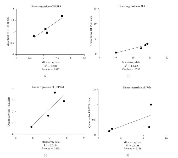
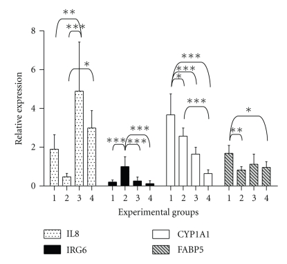
IPEC-J2, a promising in vitro model system, is not well characterized especially on the transcriptional level, in contrast to human counterparts. The aim of this study was to characterize the gene expression in IPEC-J2 cells when coincubated with enterotoxigenic Escherichia coli (ETEC), nonpathogenic E. coli, and E. coli endotoxin. Apical infection of polarized IPEC-J2 monolayers caused a time-dependent decrease in transepithelial electrical resistance (TEER). Microarray analysis showed up-regulation of interleukins when IPEC-J2 were cocultured with E. coli strains this has so far never been measured in this cell line. Highest IL8 expression was found with the ETEC strain possessing the F4 fimbrium, suggesting IPEC-J2 cells to be F4 receptor positive, confirmed in a brush border membrane adhesion assay. It is concluded that the innate immune responses to pathogens and LPS makes the IPEC-J2 cell line a suitable model for research on intestinal host pathogen interaction.
Figures
Similar articles
-
Effect of different feed ingredients and additives on IPEC-J2 cells challenged with an enterotoxigenic Escherichia coli strain.Cytotechnology. 2016 Aug;68(4):1463-71. doi: 10.1007/s10616-015-9905-6. Epub 2015 Aug 15. Cytotechnology. 2016. PMID: 26275434 Free PMC article.
-
Both enzymatic and non-enzymatic properties of heat-labile enterotoxin are responsible for LT-enhanced adherence of enterotoxigenic Escherichia coli to porcine IPEC-J2 cells.Vet Microbiol. 2013 Jun 28;164(3-4):330-5. doi: 10.1016/j.vetmic.2013.02.019. Epub 2013 Feb 28. Vet Microbiol. 2013. PMID: 23517763
-
Porcine intestinal epithelial cell lines as a new in vitro model for studying adherence and pathogenesis of enterotoxigenic Escherichia coli.Vet Microbiol. 2008 Jul 27;130(1-2):191-7. doi: 10.1016/j.vetmic.2007.12.018. Epub 2008 Jan 4. Vet Microbiol. 2008. PMID: 18261863
-
Porcine IPEC-J2 intestinal epithelial cells in microbiological investigations.Vet Microbiol. 2012 May 4;156(3-4):229-37. doi: 10.1016/j.vetmic.2011.10.017. Epub 2011 Oct 20. Vet Microbiol. 2012. PMID: 22074860 Free PMC article. Review.
-
The IPEC-J2 Cell Line.In: Verhoeckx K, Cotter P, López-Expósito I, Kleiveland C, Lea T, Mackie A, Requena T, Swiatecka D, Wichers H, editors. The Impact of Food Bioactives on Health: in vitro and ex vivo models [Internet]. Cham (CH): Springer; 2015. Chapter 12. In: Verhoeckx K, Cotter P, López-Expósito I, Kleiveland C, Lea T, Mackie A, Requena T, Swiatecka D, Wichers H, editors. The Impact of Food Bioactives on Health: in vitro and ex vivo models [Internet]. Cham (CH): Springer; 2015. Chapter 12. PMID: 29787056 Free Books & Documents. Review.
Cited by
-
Enterotoxigenic Escherichia coli CS21 pilus contributes to adhesion to intestinal cells and to pathogenesis under in vivo conditions.Microbiology (Reading). 2013 Aug;159(Pt 8):1725-1735. doi: 10.1099/mic.0.065532-0. Epub 2013 Jun 12. Microbiology (Reading). 2013. PMID: 23760820 Free PMC article.
-
Effects of the Probiotic Enterococcus faecium and Pathogenic Escherichia coli Strains in a Pig and Human Epithelial Intestinal Cell Model.Scientifica (Cairo). 2015;2015:235184. doi: 10.1155/2015/235184. Epub 2015 Mar 26. Scientifica (Cairo). 2015. PMID: 25883829 Free PMC article.
-
Differential gene expression profiling of porcine epithelial cells infected with three enterotoxigenic Escherichia coli strains.BMC Genomics. 2012 Jul 23;13:330. doi: 10.1186/1471-2164-13-330. BMC Genomics. 2012. PMID: 22823589 Free PMC article.
-
Enterococcus faecium NCIMB 10415 modulates epithelial integrity, heat shock protein, and proinflammatory cytokine response in intestinal cells.Mediators Inflamm. 2015;2015:304149. doi: 10.1155/2015/304149. Epub 2015 Apr 8. Mediators Inflamm. 2015. PMID: 25948884 Free PMC article.
-
Effects of Bacillus licheniformis and Bacillus subtilis on Gut Barrier Function, Proinflammatory Response, ROS Production and Pathogen Inhibition Properties in IPEC-J2-Escherichia coli/Salmonella Typhimurium Co-Culture.Microorganisms. 2022 Apr 29;10(5):936. doi: 10.3390/microorganisms10050936. Microorganisms. 2022. PMID: 35630380 Free PMC article.
References
-
- Hidalgo IJ, Raub TJ, Borchardt RT. Characterization of the human colon carcinoma cell line (Caco-2) as a model system for intestinal epithelial permeability. Gastroenterology. 1989;96(3):736–749. - PubMed
-
- Perlmutter DH, Daniels JD, Auerbach HS, De Schryver-Kecskemeti K, Winter HS, Alpers DH. The α-antitrypsin gene is expressed in a human intestinal epithelial cell line. Journal of Biological Chemistry. 1989;264(16):9485–9490. - PubMed
-
- Pinto M, Robine Leon S, Appay MD. Enterocyte-like differentiation and polarization of the human colon carcinoma cell line Caco-2 in culture. Biology of the Cell. 1983;47(3):323–330.
-
- Berschneider M. Development of normal cultured small intestinal epithelial cell lines which transport Na and Cl. Gastroenterology. 1989;A41
-
- Schierack P, Nordhoff M, Pollmann M, et al. Characterization of a porcine intestinal epithelial cell line for in vitro studies of microbial pathogenesis in swine. Histochemistry and Cell Biology. 2006;125(3):293–305. - PubMed
LinkOut - more resources
Full Text Sources
