. 2011 Mar 10;471(7337):220-4.
doi: 10.1038/nature09849. Epub 2011 Feb 9.

Co-adjuvant effects of retinoic acid and IL-15 induce inflammatory immunity to dietary antigens

Affiliations

Co-adjuvant effects of retinoic acid and IL-15 induce inflammatory immunity to dietary antigens

R W DePaolo et al. Nature. .

Abstract

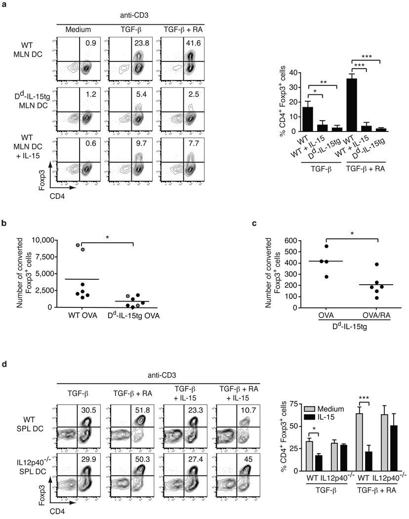
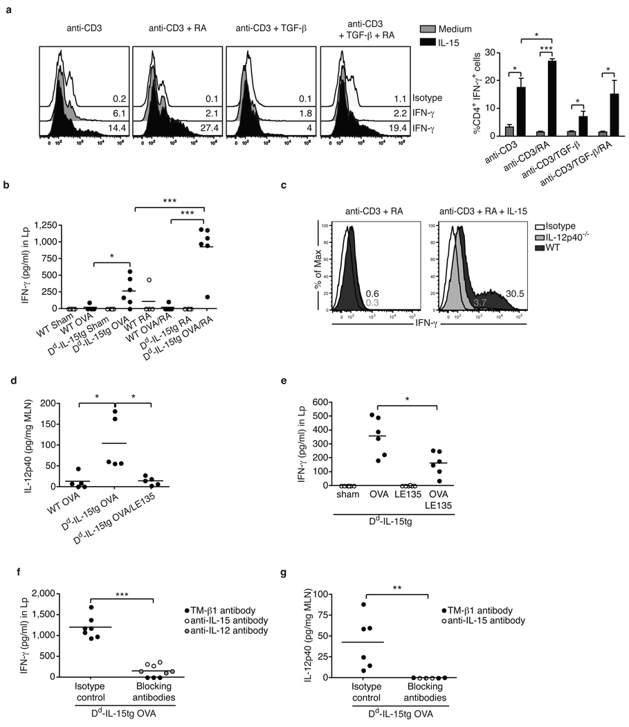
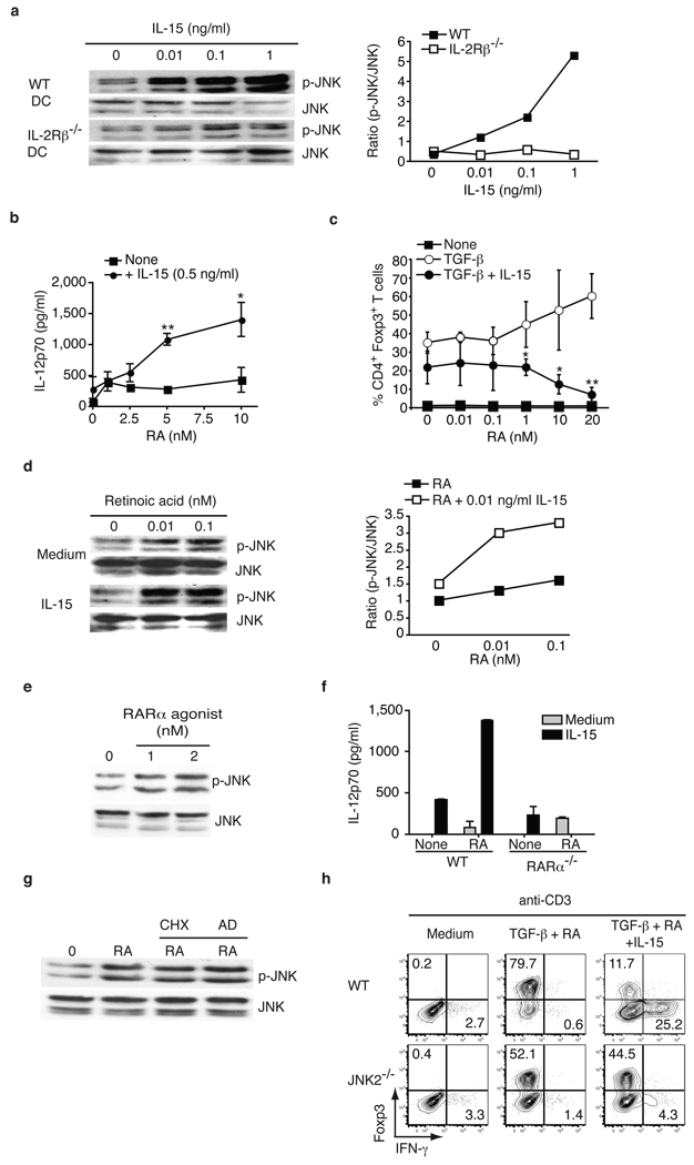
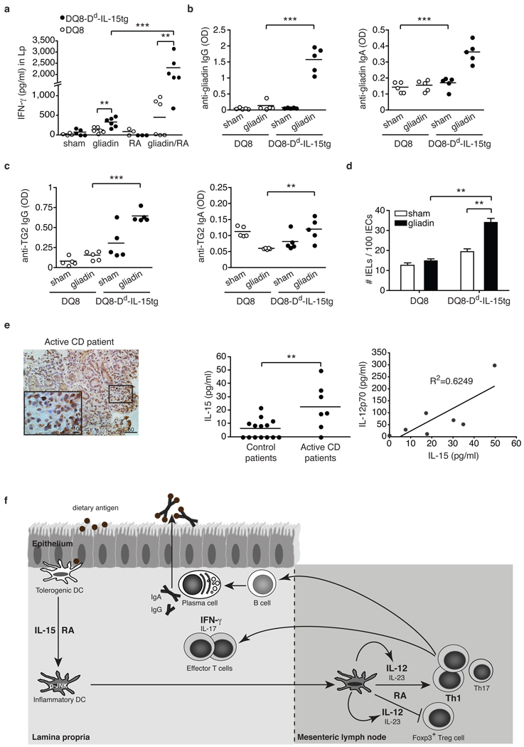
Under physiological conditions the gut-associated lymphoid tissues not only prevent the induction of a local inflammatory immune response, but also induce systemic tolerance to fed antigens. A notable exception is coeliac disease, where genetically susceptible individuals expressing human leukocyte antigen (HLA) HLA-DQ2 or HLA-DQ8 molecules develop inflammatory T-cell and antibody responses against dietary gluten, a protein present in wheat. The mechanisms underlying this dysregulated mucosal immune response to a soluble antigen have not been identified. Retinoic acid, a metabolite of vitamin A, has been shown to have a critical role in the induction of intestinal regulatory responses. Here we find in mice that in conjunction with IL-15, a cytokine greatly upregulated in the gut of coeliac disease patients, retinoic acid rapidly activates dendritic cells to induce JNK (also known as MAPK8) phosphorylation and release the proinflammatory cytokines IL-12p70 and IL-23. As a result, in a stressed intestinal environment, retinoic acid acted as an adjuvant that promoted rather than prevented inflammatory cellular and humoral responses to fed antigen. Altogether, these findings reveal an unexpected role for retinoic acid and IL-15 in the abrogation of tolerance to dietary antigens.


Figures

Figure 1
Figure 1. IL-15-activated DC in the presence of retinoic acid prevent induction of Foxp3+ regulatory T cells
a, 105 CD4+ Foxp3 T cells were cultured with 4 × 10⁴ MLN DC isolated from WT or Dd-IL-15tg mice with anti-CD3 alone or combined with IL-15, TGF-β and RA. The percentages of Foxp3+ cells are shown. Graph depicts pooled data ± s.e.m. (n=3). b, RAG1−/− OT-II CD45 congenic CD25 CD4+T cells were transferred into WT and Dd-IL-15tg mice that were fed OVA in drinking water for five days (black dots) or by gavage (grey dots). Treg cell conversion was assessed in the MLN by intracellular staining for Foxp3 and detected by flow cytometry. The absolute numbers of converted CD4+ Foxp3+ T cells are shown. Data are representative of two experiments performed independently. c, Ly5.2+ OT-II T cells were transferred into Ly5.1+ and Dd-IL-15tg-Ly5.1+ recipient mice that were fed OVA or OVA and RA five times during ten days. The absolute number of CD4+ Foxp3+ Ly5.2+ converted T cells in the MLN is shown as in a. The decrease in the number of converted iTreg was associated with a significant decrease in the number of detectable transferred T cells in Dd-IL-15tg mice (data not shown). This is likely due to the inability to detect inflammatory T cells that are more susceptible to cell death than Foxp3+ Tregs, which express anti-apoptotic factors. d, As in a, CD4+ Foxp3 T cells were cultured with SPL DC isolated from WT and IL-12p40−/− mice. The percentages of Foxp3+ cells are indicated. Graph depicts three pooled experiments ± s.e.m. *P<0.05, **P<0.01, ***P<0.001 (unpaired Student's t-test).
Figure 2
Figure 2. Retinoic acid exerts an adjuvant effect on IL-15-mediated inflammatory T cell responses
a, CD4+ T cells were cultured with WT SPL DC with the indicated cytokines. Representative histograms gated on CD4+ T cells show IFN-γ expression. The bar graph summarizes the percentage of IFN-γ-producing CD4+ T cells ± s.e.m. (n=5). b, Dd-IL-15tg and WT mice were fed PBS (sham), OVA, RA, or a mixture of OVA and RA. IFN-γ secretion by Lp cells re-stimulated for 24 h with OVA. The results are the means of triplicate samples obtained from two independent experiments. c, CD4+ T cells were cultured with SPL DC isolated from WT or IL-12p40−/− mice as described in a. Intracellular staining for IFN-γ of gated CD4 T cells is shown. Results are representative of two experiments. d, Levels of IL-12p40 in the MLN of WT and Dd-IL-15tg mice fed OVA, or a mixture of OVA and the RAR antagonist LE135. The results are the means of triplicate samples obtained from two independent experiments. Similar results were obtained for IL-12p70 and IL-23 (data not shown). e, IFN-γ secretion by Lp cells isolated from Dd-IL-15tg mice fed PBS (sham), OVA, and LE135. The results are the means of triplicate samples obtained from two independent experiments. f,g, Dd-IL-15tg mice were fed OVA and treated with blocking anti-IL-12p40, anti-IL-15, and TMβ-1 (anti-IL-2Rβ) or isotype control mAbs. The levels of IL-12p40 in the MLN (f) and IFN-γ in Lp cells re-stimulated overnight with OVA (g) were quantified. When anti-IL-15 and anti-IL-12 treatment experiments were performed in parallel, control mice received a mixture of corresponding isotype controls. Data represent two pooled experiments (n=6 mice per group) except for the anti-IL-12 treatment (n=3 individual mice). *P<0.05, ** P <0.01, *** P <0.001 (unpaired Student's t-test).
Figure 3
Figure 3. Retinoic acid and IL-15 act in synergy to induce DC with proinflammatory properties in a JNK-dependent manner
a, Concentration-dependent JNK phosphorylation in WT or IL-2Rβ−/− BMDC upon IL-15 stimulation analyzed by western blot (left panel) and quantified (right panel). b, IL-12p70 secretion after overnight culture of WT SPL DC with increasing doses of RA, with and without IL-15. Results are mean values ± s.e.m. (n=3). c, CD4+ Foxp3 T cells were cultured with WT SPL DC with anti-CD3 alone or combined with IL-15, TGF-β and increasing doses of RA. The percentages of Foxp3+ cells are shown. Graph depicts pooled data ± s.e.m (n=3). d, Concentration-dependent JNK phosphorylation in WT BMDC upon IL-15 (0.01 ng/ml) and increasing doses of RA stimulation by western blot (left panel) and quantified (right panel). e, Concentration-dependent JNK phosphorylation in WT BMDC upon stimulation with a RARα agonist (AM580). f, IL-12p70 secretion after overnight culture of WT and RARα−/− BMDC with IL-15 alone or combined with RA. Data are shown as means and s.e.m. (n=2). g, JNK phosphorylation in BMDC pretreated with cyclohexamide (CHX) or actinomycin D (AD) prior to stimulation with 0.1 nM RA. h, CD4+ Foxp3 T cells were cultured with SPL DC isolated from WT or JNK2−/− mice with the indicated cytokines. The percentages of Foxp3+ and IFN-γ+ among CD4+ T cells are indicated. Results are representative of two independent experiments. *P<0.05, ** P <0.01, (unpaired Student's t-test).
Figure 4
Figure 4. DQ8-Dd-IL-15tg mice fed gliadin mimic early stages of celiac disease reflecting dysregulation in the adaptive immune response to gluten
a-d, DQ8 and DQ8-Dd-IL-15tg mice were fed gliadin every other day for ten days. a, IFN-γ secretion by Lp cells after overnight culture with gliadin. b, c, Anti-gliadin IgG, anti-gliadin IgA, anti-TG2 IgG and anti-TG2 IgA titers from serum collected fifteen days after feeding. d, Quantification of IEL among intraepithelial cells in small intestines fifteen days after the last feeding. e, IL-15 and IL-12 expression in the Lp of CD patients. Immunohistochemical stainings for IL-15 in gut tissue from an active celiac disease patient (left panel). Lp cells were harvested from biopsies obtained from control (n=14) or active CD patients (n=7) and assayed for levels of IL-15 and IL-12p70 by ELISA (middle and right panels). Equal concentration of total proteins was analyzed for each sample. **P<0.01, ***P<0.001 (unpaired Student's t-test). f, Proposed model for the co-adjuvant effects of RA and IL-15 in the intestinal mucosa. Under inflammatory conditions, the expression of the pro-inflammatory cytokine IL-15 is upregulated in the Lp of the small intestine. Through the synergistic action of IL-15 and RA, DC acquire the ability to release inflammatory cytokines, particularly IL-12 and IL-23. These inflammatory mediators then act in concert with RA to prevent the induction of Foxp3+ Treg cells and drive TH1 and potentially TH17 polarization. In turn, inflammatory T cells may provide help to B cells to produce specific IgG and IgA antibodies.

Comment in

Similar articles

  • Immunology: Context is key in the gut.
    Maynard CL, Weaver CT. Maynard CL, et al. Nature. 2011 Mar 10;471(7337):169-70. doi: 10.1038/471169a. Nature. 2011. PMID: 21390118 No abstract available.
  • Gliadin Nanoparticles Induce Immune Tolerance to Gliadin in Mouse Models of Celiac Disease.
    Freitag TL, Podojil JR, Pearson RM, Fokta FJ, Sahl C, Messing M, Andersson LC, Leskinen K, Saavalainen P, Hoover LI, Huang K, Phippard D, Maleki S, King NJC, Shea LD, Miller SD, Meri SK, Getts DR. Freitag TL, et al. Gastroenterology. 2020 May;158(6):1667-1681.e12. doi: 10.1053/j.gastro.2020.01.045. Epub 2020 Feb 4. Gastroenterology. 2020. PMID: 32032584 Free PMC article.
  • IL-15, gluten and HLA-DQ8 drive tissue destruction in coeliac disease.
    Abadie V, Kim SM, Lejeune T, Palanski BA, Ernest JD, Tastet O, Voisine J, Discepolo V, Marietta EV, Hawash MBF, Ciszewski C, Bouziat R, Panigrahi K, Horwath I, Zurenski MA, Lawrence I, Dumaine A, Yotova V, Grenier JC, Murray JA, Khosla C, Barreiro LB, Jabri B. Abadie V, et al. Nature. 2020 Feb;578(7796):600-604. doi: 10.1038/s41586-020-2003-8. Epub 2020 Feb 12. Nature. 2020. PMID: 32051586 Free PMC article.
  • Pathomechanisms in celiac disease.
    Dieterich W, Esslinger B, Schuppan D. Dieterich W, et al. Int Arch Allergy Immunol. 2003 Oct;132(2):98-108. doi: 10.1159/000073710. Int Arch Allergy Immunol. 2003. PMID: 14600421 Review.
  • Celiac disease: how complicated can it get?
    Tjon JM, van Bergen J, Koning F. Tjon JM, et al. Immunogenetics. 2010 Oct;62(10):641-51. doi: 10.1007/s00251-010-0465-9. Epub 2010 Jul 27. Immunogenetics. 2010. PMID: 20661732 Free PMC article. Review.

Cited by

References

    1. Curotto de Lafaille MA, Lafaille JJ. Natural and adaptive foxp3+ regulatory T cells: more of the same or a division of labor? Immunity. 2009;30(5):626. - PubMed
    1. Faria AM, Weiner HL. Oral tolerance. Immunol Rev. 2005;206:232. - PMC - PubMed
    1. Jabri B, Sollid LM. Tissue-mediated control of immunopathology in coeliac disease. Nat Rev Immunol. 2009;9(12):858. - PubMed
    1. Mora JR, Iwata M, von Andrian UH. Vitamin effects on the immune system: vitamins A and D take centre stage. Nat Rev Immunol. 2008 - PMC - PubMed
    1. Coombes JL, et al. A functionally specialized population of mucosal CD103+ DCs induces Foxp3+ regulatory T cells via a TGF-beta and retinoic acid-dependent mechanism. J Exp Med. 2007;204(8):1757. - PMC - PubMed

Publication types

MeSH terms