. 2011 Mar 1;108(9):3624-9.
doi: 10.1073/pnas.1016201108. Epub 2011 Feb 7.

Endogenous retrovirus drives hitherto unknown proapoptotic p63 isoforms in the male germ line of humans and great apes

Affiliations

Endogenous retrovirus drives hitherto unknown proapoptotic p63 isoforms in the male germ line of humans and great apes

Ulrike Beyer et al. Proc Natl Acad Sci U S A. .

Abstract

TAp63, but not its homolog p53, eliminates oocytes that suffered DNA damage. An equivalent gene for guarding the male germ line is currently not known. Here we identify hitherto unknown human p63 transcripts with unique 5'-ends derived from incorporated exons upstream of the currently mapped TP63 gene. These unique p63 transcripts are highly and specifically expressed in testis. Their most upstream region corresponds to a LTR of the human endogenous retrovirus 9 (ERV9). The insertion of this LTR upstream of the TP63 locus occurred only recently in evolution and is unique to humans and great apes (Hominidae). A corresponding p63 protein is the sole p63 species in healthy human testis, and is strongly expressed in spermatogenic precursors but not in mature spermatozoa. In response to DNA damage, this human male germ-cell-encoded TAp63 protein (designated GTAp63) is activated by caspase cleavage near its carboxyterminal domain and induces apoptosis. Human testicular cancer tissues and cell lines largely lost p63 expression. However, pharmacological inhibition of histone deacetylases completely restores p63 expression in testicular cancer cells (>3,000-fold increase). Our data support a model whereby testis-specific GTAp63 protects the genomic integrity of the male germ line and acts as a tumor suppressor. In Hominidae, this guardian function was greatly enhanced by integration of an endogenous retrovirus upstream of the TP63 locus that occurred 15 million years ago. By providing increased germ-line stability, this event may have contributed to the evolution of hominids and enabled their long reproductive periods.


Conflict of interest statement

The authors declare no conflict of interest.

Figures
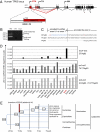
Fig. 1.
Fig. 1.
Testis-specific p63 isoforms are encoded by unique upstream exons, as a result of recent retroviral LTR insertion. (A) Genomic architecture of the human TP63 gene, including all known (black and gray) and previously unexplored (red) exons. The recently identified upstream exons U1, U2, and U3 encode GTAp63. PremRNA splicing fuses these exons directly to the previously described exon 2, thus omitting exon 1. Predominant splicing is indicated by solid lines, less abundant splicing patterns by dotted lines. The three start sites of transcription (GTA, TA, and ΔN) are indicated with arrows above their first exons. Alternative splicing at the downstream exons 10 to 14 generates isoforms designated α (the major variant), β, and γ. (Inset) ERV9 LTR overlaps with the first upstream exon U1 and contains the transcriptional start site (TSS) of GTAp63. (B) Expression of ERV9 LTR-driven p63 transcripts in human testis. Human testicular cDNA was amplified with primers binding to exon U1 and exon 2, as indicated in Fig. S1A and Table S1. The size of the predominantly amplified PCR product corresponds to exon U1 directly spliced onto exon 2. An additional, less-abundant transcript also contains exon U2. (C) Predicted amino acid sequence of human GTAp63 derived from the major transcript present in testis (B). Note that GTAp63 differs from the previously described, long TAp63, termed TA*p63 (2), by a unique 19-residue long amino terminus. TAp63 is a shorter isoform because it uses the second ATG within exon 2 as start codon (2). (D) The unique GTAp63 transcripts are highly expressed in testis, but barely or not at all in somatic tissues. GTAp63 also shows some expression in the ovary of a young female donor but much less so from an older woman (see for example, Fig. S1E). Relative mRNA levels of GTAp63 and conventional TAp63 in an array of normal human tissues were determined by quantitative RT-PCR. Human testis contained 500- to 1,000-fold more GTAp63 transcripts than any of the few somatic tissues with detectable expression (note the logarithmic scale). Most tissues do not express detectable GTAp63. (E) Timing of primate evolution and ERV9 insertion adjacent to TP63. The pattern and sequence of TP63-associated ERV9 LTR in primate genomes (Fig. S1G) indicates that the insertion of the ERV9 LTR upstream of TP63 occurred during the time shortly after Hominidae separated from Hylobatidae, roughly 15 million years ago. This theory is compatible with the model that this insertion contributed to enhanced genomic stability of the male germ line, perhaps enabling the preservation of species with a much prolonged time frame of fertility and increased complexity in gene regulation.
Fig. 2.
Fig. 2.
High levels of GTAp63 in human spermatogonia. (A) Human testis expresses high levels of GTAp63 protein as the sole p63 species, as revealed by immunoblot analysis. The band detected by the p63 antibody 4A4 in human testis but not in liver or ovarian tissue corresponds to recombinant GTAp63. Overexpression of GTAp63 in H1299 cells. Nonspecific cross-reactive bands are labeled (asterisk). (B) Expression of p63 in normal human testis. Two representative examples of seminiferous tubules were immunostained with a polyclonal antibody to p63 (# 9424). Although all layers of precursors except the mature luminal spermatozoa (asterisk) stain positively, the highest level of p63 tends to be present in the mitotic spermatogonia at the base (single arrowheads). Suprabasal, meiotic spermatozytes (double arrowheads) tend to express less p63. (C) A similar pattern of p63 expression in normal human testis is seen with monoclonal p63 antibody 4A4. For a lower magnification, see Fig. S2A. (D) Expression of p63 in normal murine testis. Testicular tissue from a 4-mo-old SV129 mouse was immunostained with monoclonal p63 antibody 4A4. Basal spermatogonia reveal only a weak signal (single arrowheads), although the maximum staining intensity is shifted to suprabasal spermatocytes (double arrowheads). Mature luminal spermatozoa (asterisk) are completely negative. For a control staining, see Fig. S2B. The absence of a specific signal upon staining of p63−/− testis with antibody 4A4 is documented in Fig. S2C.
Fig. 3.
Fig. 3.
GTAp63 induces apoptosis in response to genotoxic stress. (A) Schematic of the GTAp63α domain structure, including the DNA binding domain (DBD), the oligomerization domain (oligo), and the SAM domain. Point mutations that were introduced for functional analysis are indicated. Mutation C306R disrupts DNA binding (28), whereas mutation D497A prevents caspase cleavage (20). A stop codon at position 498 results in the synthesis of a truncated, constitutively active GTAp63 that mimics the fragment generated by caspase cleavage in response to DNA damage (used in Fig. S3C). Note, the numbering of positions refers to conventional TAp63, starting at the second ATG (see Fig. 1C, TAp63), to match the published numbering (28). (B) In response to cisplatin treatment, caspase-cleavable wild-type GTAp63 decreases long-term clonogenic survival. H1299 cells (p53 null) were stably transfected with empty vector (pIRESneo, Clontech; control) or expression constructs for wild-type GTAp63α or its mutants. The cells were treated with cisplatin for 24 h, followed by drug removal and culturing for 7 d. Emerging clones were stained with Crystal violet. (C) Quantitation of the plate area covered by cell clones from B. Columns represent mean values and the SEM. P values were calculated using Student's t test. (D) Apoptosis in response to DNA damage depends on GTAp63. H1299 cells stably expressing GTAp63 or its mutants were treated with cisplatin for 24 h. Activation of caspases was detected by staining with fluorescently labeled DEVD, indicating mid- and late-stage apoptosis. The mean proportion of apoptotic cells and its SE are indicated. The corresponding raw data are shown in Fig. S3B. (E) Induction of proapoptotic genes by wild-type GTAp63 in response to cisplatin. GTAp63α or the indicated mutants were stably expressed in H1299 cells. Cells were treated with cisplatin or DMSO for 24 h, followed by quantitative RT-PCR to determine the levels of p53/p63-responsive genes. Wild-type GTAp63 induces the proapoptotic genes PUMA/BBC3, Noxa/PMAIP1, and CD95L/TNF6SF6, but DNA-binding and noncleavable mutants do not. On the other hand, the cell-cycle regulatory genes CDKN1A/p21 and GADD45A are not or only minimally induced by GTAp63. P values were calculated as described in C.
Fig. 4.
Fig. 4.
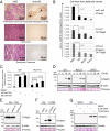
GTAp63 is frequently lost in testicular germ-cell cancers, but expression can be fully rescued by HDAC inhibitors. (A) Frequent loss of p63 expression in invasive human testicular germ cell cancers. Representative examples of seminomas and embryonal carcinomas (the two most important testicular cancer types) are shown. Staining with H&E and polyclonal p63 antibody 9424, as in Fig. 2B. The faint cellular outlines are nonspecific. More examples are in Fig. S4A. The results obtained from all 98 tumors are summarized in Table 1. (Upper) In contrast, preinvasive germ-cell intratubular neoplasms (GCIN) mostly preserve expression of nuclear p63 (arrowheads). Seminiferous tubules are completely filled with atypical GCIN cells that lack normal differentiation and spermatogenesis. (Left) Large atypical tumor cells arranged in sheets separated by loose fibrous stroma. H&E staining. (Right) Shows that p63 is lost. (B) Compared with normal human testis, GTAp63 mRNA expression is strongly reduced in all three randomly chosen cell lines from human testicular germ-cell cancers. P values were calculated as described in Fig. 3C. (C) GTAp63 is lost in testicular cancer cell lines via transcriptional silencing but fully restored by HDAC inhibition. Real-time RT-PCR to quantify GTAp63 mRNA, normalized for GAPDH. GH cells (teratocarcinoma) were treated with TSA for 24 h. This treatment increased the level of GTAp63 mRNA up to 3,000-fold (logarithmic scale). No comparable increase was seen for conventional TAp63 or TAp73 in the same cells (Fig. S4B). Moreover, only a very modest increase of GTAp63 (20-fold) was seen in colon carcinoma cells (HCT116). P values were calculated as described in Fig. 3C. (D) Restoration of p63 protein in human testicular cancer cells upon HDAC inhibition. GH and NCCIT cells (teratocarcinomas) were treated with 500 nM TSA, followed by 4A4 immunoblot detection of p63. In contrast, U2OS human osteosarcoma cells treated in parallel exhibit no detectable GTAp63. (E) Silencing of p63 via siRNA abolishes the GTAp63 protein detectable in TSA-treated GH cells, confirming its identity. (F) The single band detectable by anti-p63 antibodies in TSA-treated GH cells corresponds to the molecular weight of recombinant GTAp63 expressed in H1299 cells (a lung adenocarcinoma line with no detectable endogenous p63). (G) SAHA also induces GTAp63 protein in testicular cancer cells but not in osteosarcoma cells. GH cells (teratocarcinoma) and U2OS cells (osteosarcoma) were treated with SAHA for 24 h, followed by immunoblot detection of p63. A band corresponding to the molecular weight of recombinant GTAp63 was only detected in SAHA-treated GH cells.

Similar articles

Cited by

References

    1. Arnheim N, Calabrese P. Understanding what determines the frequency and pattern of human germline mutations. Nat Rev Genet. 2009;10:478–488. - PMC - PubMed
    1. Yang A, et al. p63, a p53 homolog at 3q27-29, encodes multiple products with transactivating, death-inducing, and dominant-negative activities. Mol Cell. 1998;2:305–316. - PubMed
    1. Yang A, McKeon F. P63 and P73: P53 mimics, menaces and more. Nat Rev Mol Cell Biol. 2000;1:199–207. - PubMed
    1. Lane DP. Cancer. p53, guardian of the genome. Nature. 1992;358:15–16. - PubMed
    1. Vousden KH, Prives C. Blinded by the light: The growing complexity of p53. Cell. 2009;137:413–431. - PubMed

Publication types

MeSH terms