. 2011 Mar 25;286(12):10495-504.
doi: 10.1074/jbc.M110.195628. Epub 2011 Feb 2.

Pancreatic cancer cells respond to type I collagen by inducing snail expression to promote membrane type 1 matrix metalloproteinase-dependent collagen invasion

Affiliations

Pancreatic cancer cells respond to type I collagen by inducing snail expression to promote membrane type 1 matrix metalloproteinase-dependent collagen invasion

Mario A Shields et al. J Biol Chem. .

Abstract

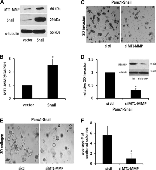
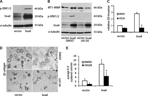
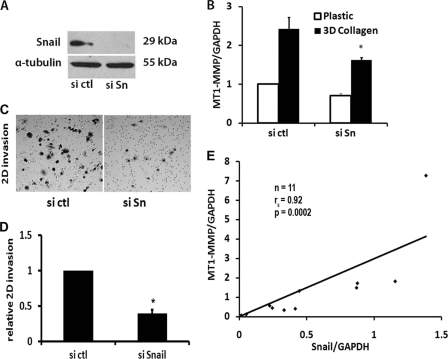
Pancreatic ductal adenocarcinoma (PDAC) is characterized by pronounced fibrotic reaction composed primarily of type I collagen. Although type I collagen functions as a barrier to invasion, pancreatic cancer cells have been shown to respond to type I collagen by becoming more motile and invasive. Because epithelial-mesenchymal transition is also associated with cancer invasion, we examined the extent to which collagen modulated the expression of Snail, a well known regulator of epithelial-mesenchymal transition. Relative to cells grown on tissue culture plastic, PDAC cells grown in three-dimensional collagen gels induced Snail. Inhibiting the activity or expression of the TGF-β type I receptor abrogated collagen-induced Snail. Downstream of the receptor, we showed that Smad3 and Smad4 were critical for the induction of Snail by collagen. In contrast, Smad2 or ERK1/2 was not involved in collagen-mediated Snail expression. Overexpression of Snail in PDAC cells resulted in a robust membrane type 1-matrix metalloproteinase (MT1-MMP, MMP-14)-dependent invasion through collagen-coated transwell chambers. Snail-expressing PDAC cells also demonstrated MT1-MMP-dependent scattering in three-dimensional collagen gels. Mechanistically, Snail increased the expression of MT1-MMP through activation of ERK-MAPK signaling, and inhibiting ERK signaling in Snail-expressing cells blocked two-dimensional collagen invasion and attenuated scattering in three-dimensional collagen. To provide in vivo support for our findings that Snail can regulate MT1-MMP, we examined the expression of Snail and MT1-MMP in human PDAC tumors and found a statistically significant positive correlation between MT1-MMP and Snail in these tumors. Overall, our data demonstrate that pancreatic cancer cells increase Snail on encountering collagen-rich milieu and suggest that the desmoplastic reaction actively contributes to PDAC progression.


Figures

FIGURE 1.
FIGURE 1.
Collagen-induced Snail expression is mediated by TGF-β signaling. A, Panc1 and HPDE cells were grown on tissue culture plastic or in three-dimensional type I collagen gels (2 mg/ml) for 24 h, and Snail mRNA expression was analyzed by qRT-PCR using the Comparative CT method. The -fold change in Snail expression relative to plastic was determined using GAPDH as normalization control. *, p < 0.05. B, Panc1 cells were plated on tissue culture plastic, serum-starved overnight, then treated with TGF-β1 (10 ng/ml) for 12 h. The cell lysates were analyzed for Snail protein expression by Western blot analysis using α-tubulin as normalization control. C, HPDE and Panc1 cells were grown in three-dimensional collagen in the presence of TβRI kinase inhibitor SB431542 (SB, 10 μm) or DMSO (DM, vehicle control) for 24 h, and then the expression of Snail was analyzed by qRT-PCR. *, p < 0.05. D, Panc1 cells were transfected with a control siRNA (si ctl) or TβRI specific siRNA (si TβRI), allowed to recover, and after 48 h plated on tissue culture plastic or in three-dimensional collagen gels for 24 h. The knockdown of the TβRI protein was analyzed by Western blot analysis using α-tubulin as normalization control (top). The effect of TβRI knockdown on collagen-induced Snail expression was examined using qRT-PCR (bottom). The results are representative of at least three independent experiments.
FIGURE 2.
FIGURE 2.
Smad3, but not Smad2, regulates collagen-induced Snail expression. A, Panc1 cells were plated on tissue culture plastic (PL) or in three-dimensional collagen gels (2 mg/ml, COL) for 24 h in the presence of DMSO or U0126 (10 μm). The lysates were then analyzed for ERK1/2 phosphorylation and α-tubulin by Western blot analysis (top). The cell lysates were also analyzed for Snail expression by qRT-PCR using GAPDH as normalization control (bottom). B–D, Panc1 cells were transfected with control siRNA (si ctl), siRNA against Smad4 (si Sd4, B), Smad2 (si Sd2, C) or Smad3 (si Sd3, D) (50 nm). After 48 h the transfected cells were plated on plastic or in three-dimensional collagen for 24 h. The specific down-regulation of each of the Smad proteins was analyzed by Western blot analysis using α-tubulin as normalization control (top). The effect of knockdown of each of the Smad on collagen-induced Snail mRNA expression was analyzed by qRT-PCR using GAPDH as normalization control (bottom). *, p < 0.05. The results are representative of at least three independent experiments.
FIGURE 3.
FIGURE 3.
Snail promotes MMP-dependent two-dimensional collagen invasion and induces MMP-dependent scattering in three-dimensional collagen gels. A, Panc1 cells were transfected with pTet-On (Clontech) vector and co-transfected with pTight-Luc or pTight-Snail and cell lines resistant to both G418 and puromycin selected (Panc1-vector, Panc1-Snail). Equal number of Panc1-vector and Panc1-Snail cells were plated on plastic and treated with or without doxycycline (2 μg/ml) for 16 h. The expression of Snail was analyzed by Western blot analysis using α-tubulin as normalization control. B and C, Panc1-vector and Panc1-Snail cells (2 × 10⁵) were added in 800 μl of serum-free medium to the upper chamber of BD Biocoat porous polycarbonate filters (8 μm pore) coated with 7.5 μg of type I collagen, and 1 ml of serum-containing medium supplemented with doxycycline was added to the lower chamber. The cells were allowed to invade over 30 h, and the non-invading cells were removed from the upper chamber. The filters were then fixed, stained, and photographed (B), and the relative invasion was quantified (C). D and E, Panc1-vector and Panc1-Snail cells were embedded in three-dimensional collagen gels (2 mg/ml), and doxycycline-containing media was changed every 2 days for 6 days. The effect of Snail on colony morphology was examined by phase contrast microscopy (D), and the number of scattered colonies per field was quantified as detailed under “Experimental Procedures” (E). F and G, Panc1-vector and Panc1-Snail cells were grown in three-dimensional collagen gels (2 mg/ml) and doxycycline with DMSO (vehicle control) or GM6001 (10 μm) added every 2 days for 6 days. The effect on colony morphology in at least five fields was examined by phase contrast microscopy (F), and the average number of scattered colonies per field was quantified (G). H, the effect of GM6001 on invasion of Panc1-vector and Panc1-Snail cells through type I collagen-coated membrane was also determined. *, p < 0.05. The results are representative of at least three independent experiments.
FIGURE 4.
FIGURE 4.
Snail increases MT1-MMP expression in pancreatic cancer cells. A and B, Panc1- vector and Panc1-Snail cells were induced with doxycycline (2 μg/ml) for 24 h, and the lysates were analyzed for Snail, MT1-MMP, and α-tubulin protein expression by Western blotting (A) and MT1-MMP and GAPDH mRNA expression by qRT-PCR (B). C and D, doxycycline-treated Panc1-Snail cells were transfected with control siRNA (si ctl) or MT1-MMP-specific siRNA (si MT1-MMP), allowed to recover for 24 h, and added to type I collagen-coated transwell chambers as detailed in Fig. 3 and allowed to invade. The knock down of MT1-MMP was determined after 48 h by Western blotting using α-tubulin as loading control (D, inset). A representative figure of the invading cells after 24 h (C) and the quantification of the relative invasion from 4 independent experiments are shown (D). E and F, the transfected Panc1-Snail cells were also seeded in three-dimensional collagen gels (2 mg/ml), doxycycline-containing media supplemented with control siRNA or MT1-MMP-specific siRNA was changed every 2 days for 6 days, the effect on colony morphology was examined by phase microscopy (E), and the average number of scattered colonies was quantified (F). The results are representative of at least three independent experiments. *, p < 0.05.
FIGURE 5.
FIGURE 5.
TIMP-2 blocks Snail-induced scattering in three-dimensional. A and B, Panc1-Snail cells were grown in three-dimensional collagen gels (2 mg/ml) with TIMP-1 (5 μg/ml) or TIMP-2 (5 μg/ml)-co-polymerized. The doxycycline-containing media supplemented with TIMP-1 or TIMP-2 was changed every 2 days for 6 days, the effect on colony morphology was examined by phase microscopy (A), and the average number of scattered colonies quantified (B). *, p < 0.05. C, Panc1-Snail cells were transfected with control siRNA (si ctl) or MMP-2-specific siRNA (si MMP2) and allowed to recover for 24 h. The cells were then serum-starved for 24 h, and the conditioned media were analyzed for MMP-2 expression by gelatin zymography. D and E, the transfected Panc1-Snail cells were also seeded in three-dimensional collagen gels (2 mg/ml), the doxycycline-containing media supplemented with control siRNA or MMP-2-specific siRNA were changed every 2 days for 6 days, the effect on colony morphology was examined by phase microscopy (D), and the average number of scattered colonies was quantified (E). The results are representative of at least three independent experiments.
FIGURE 6.
FIGURE 6.
ERK1/2 mediates Snail-induced MT1-MMP expression and collagen invasion. A, Panc1-vector and Panc1-Snail cells were induced with doxycycline (2 μg/ml) for 24 h, and the lysates were analyzed for Snail, p-ERK1/2, and α-tubulin expression by Western blot analysis. B, Panc1-vector and Panc1-Snail cells were induced with doxycycline (2 μg/ml) for 24 h in the presence of DMSO or MEK1/2 inhibitor U0126 (10 μm). The lysates were analyzed for Snail, p-ERK1/2, MT1-MMP, and α-tubulin expression by Western blotting. C, Panc1-vector and Panc1-Snail cells (2 × 10⁵) in 800 μl of serum-free medium were added to the upper chamber of BD Biocoat porous polycarbonate filters (8 μm pore) coated with 7.5 μg of type I collagen and 1 ml of serum containing medium supplemented with doxycycline, and either DMSO or U0126 (10 μm) was added to the lower chamber. The cells were allowed to invade for 30 h, and the non-invading cells were removed from the upper chamber, membranes were fixed, and the relative invasion was quantified. D and E, Panc1-vector or Panc1-Snail cells were suspended in three-dimensional gel (2 mg/ml), and fresh serum-containing medium supplemented with doxycycline and either DMSO or U0126 (10 μm) was added every 2 days for 6 days. The effect on colony morphology was examined by phase contrast microscopy (D), and the average number of scattered colonies per field was quantified (E). The results are representative of at least three independent experiments. *, p < 0.05.
FIGURE 7.
FIGURE 7.
Collagen-induced MT1-MMP expression involves Snail. A and B, Panc1 cells were transfected with control siRNA (si ctl) or a combination of two different Snail specific siRNAs (si Sn), allowed to recover for 48 h, and then plated on tissue culture plastic or in collagen gels for 24 h. The specific knockdown of Snail protein was determined by Western blotting using α-tubulin as normalization control (A), and the effect of Snail siRNA on collagen-induced MT1-MMP was analyzed by qRT-PCR using GAPDH as normalization control (B). The results are representative of at least three independent experiments. *, p < 0.05. C and D, Panc1 cells were transfected with control siRNA or specific siRNAs, allowed to recover for 24 h, added to type I collagen-coated transwell chambers as detailed in Fig. 3, and allowed to invade. A representative figure of the invading cells after 24 h (C) and the quantification of the relative invasion from 4 independent experiments are shown (D). *, p < 0.05. E, using de-identified human pancreatic tissue specimens (n = 11) obtained on an institutional review board-approved protocol, RNA was isolated from human PDAC tumors and the adjacent matched normal pancreas and analyzed by qRT-PCR. Snail and MT1-MMP expression in PDAC tumors was normalized to the levels present in the matched adjacent normal pancreas, which was arbitrarily set at 1.0, and analyzed using Spearman's correlation.

Similar articles

Cited by

References

    1. Thiery J. P., Acloque H., Huang R. Y., Nieto M. A. (2009) Cell 139, 871–890 - PubMed
    1. Kalluri R., Weinberg R. A. (2009) J. Clin. Invest. 119, 1420–1428 - PMC - PubMed
    1. Lee J. M., Dedhar S., Kalluri R., Thompson E. W. (2006) J. Cell Biol. 172, 973–981 - PMC - PubMed
    1. Batlle E., Sancho E., Francí C., Domínguez D., Monfar M., Baulida J., García De Herreros A. (2000) Nat. Cell Biol. 2, 84–89 - PubMed
    1. Cano A., Pérez-Moreno M. A., Rodrigo I., Locascio A., Blanco M. J., del Barrio M. G., Portillo F., Nieto M. A. (2000) Nat. Cell Biol. 2, 76–83 - PubMed

Publication types

MeSH terms

Substances