Slug enhances invasion ability of pancreatic cancer cells through upregulation of matrix metalloproteinase-9 and actin cytoskeleton remodeling
- PMID: 21283078
- PMCID: PMC3125102
- DOI: 10.1038/labinvest.2010.201
Slug enhances invasion ability of pancreatic cancer cells through upregulation of matrix metalloproteinase-9 and actin cytoskeleton remodeling
Retraction in
-
Retraction. Slug enhances invasion ability of pancreatic cancer cells through upregulation of matrix metalloproteinase-9 and actin cytoskeleton remodeling.Lab Invest. 2012 Dec;92(12):1801. doi: 10.1038/labinvest.2012.138. Epub 2012 Nov 19. Lab Invest. 2012. PMID: 23191991 Free PMC article. No abstract available.
Abstract
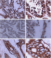
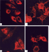
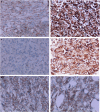
Slug, a member of the Snail family of transcription factors, has a crucial role in the regulation of epithelial-mesenchymal transition (EMT) by suppressing several epithelial markers and adhesion molecules, including E-cadherin. A recent study demonstrated that no relationship exists between Slug and E-cadherin in pancreatic cancer. Another study showed that in malignant mesothelioma effusions Slug was associated with matrix metalloproteinase (MMP) expression, but that there was no association with E-cadherin. F-ascin is an actin-bundling protein involved in filopodia assembly and cancer invasion and metastasis of multiple epithelial cancer types. In this study, we investigated Slug, E-cadherin, and MMP-9 expression using immunohistochemistry in 60 patients with pancreatic cancer and their correlation with carcinoma invasion and metastasis. Additionally, we observed the effects of Slug on invasion and metastasis in the pancreatic cancer cell line PANC-1. Alterations in Slug, MMP-9, and E-cadherin were determined by RT-PCR, western blot, and immunohistochemistry. Alterations in MMP-9 and F-actin cytoskeleton were determined by immunofluorescence staining, flow cytometry (FCM), or gelatin zymography. Slug, E-cadherin, and MMP-9 expression in pancreatic cancer was significantly associated with lymph node metastases and we found a significant correlation between Slug and MMP-9 expression; however, no significant correlation was observed between Slug and E-cadherin expression. Slug transfection significantly increased invasion and metastasis in PANC-1 cells and orthotopic tumor of mouse in vivo, and significantly upregulated and activated MMP-9; however, there was no effect on E-cadherin expression. Slug promoted the formation of lamelliopodia or filopodia in PANC-1 cells. The intracellular F-actin and MMP-9 was increased and relocated to the front of the extending pseudopodia from the perinuclear pool in Slug-transfected PANC-1 cells. These results suggest that Slug promotes migration and invasion of PANC-1 cells, which may correlate with the reorganization of MMP-9 and remodeling of the F-actin cytoskeleton, but not with E-cadherin expression.
Figures


Similar articles
-
Expression of Snail, Slug and Sip1 in malignant mesothelioma effusions is associated with matrix metalloproteinase, but not with cadherin expression.Lung Cancer. 2006 Dec;54(3):309-17. doi: 10.1016/j.lungcan.2006.08.010. Epub 2006 Sep 25. Lung Cancer. 2006. PMID: 16996643
-
Matrix metalloproteinase-9 cooperates with transcription factor Snail to induce epithelial-mesenchymal transition.Cancer Sci. 2011 Apr;102(4):815-27. doi: 10.1111/j.1349-7006.2011.01861.x. Epub 2011 Feb 9. Cancer Sci. 2011. PMID: 21219539
-
Expression of Snail and Slug in renal cell carcinoma: E-cadherin repressor Snail is associated with cancer invasion and prognosis.Lab Invest. 2011 Oct;91(10):1443-58. doi: 10.1038/labinvest.2011.111. Epub 2011 Aug 1. Lab Invest. 2011. PMID: 21808237
-
The Role of MMP-9 and MMP-9 Inhibition in Different Types of Thyroid Carcinoma.Molecules. 2023 Apr 25;28(9):3705. doi: 10.3390/molecules28093705. Molecules. 2023. PMID: 37175113 Free PMC article. Review.
-
MMP-7 marks severe pancreatic cancer and alters tumor cell signaling by proteolytic release of ectodomains.Biochem Soc Trans. 2022 Apr 29;50(2):839-851. doi: 10.1042/BST20210640. Biochem Soc Trans. 2022. PMID: 35343563 Free PMC article. Review.
Cited by
-
Manganese superoxide dismutase induces migration and invasion of tongue squamous cell carcinoma via H2O2-dependent Snail signaling.Free Radic Biol Med. 2012 Jul 1;53(1):44-50. doi: 10.1016/j.freeradbiomed.2012.04.031. Epub 2012 May 9. Free Radic Biol Med. 2012. PMID: 22580338 Free PMC article.
-
Biomarkers for predicting future metastasis of human gastrointestinal tumors.Cell Mol Life Sci. 2013 Oct;70(19):3631-56. doi: 10.1007/s00018-013-1266-8. Epub 2013 Jan 31. Cell Mol Life Sci. 2013. PMID: 23370778 Free PMC article. Review.
-
Human pancreatic adenocarcinoma contains a side population resistant to gemcitabine.BMC Cancer. 2012 Aug 15;12:354. doi: 10.1186/1471-2407-12-354. BMC Cancer. 2012. PMID: 22894607 Free PMC article.
-
Epithelial-to-mesenchymal transition in pancreatic ductal adenocarcinoma: Characterization in a 3D-cell culture model.World J Gastroenterol. 2016 May 14;22(18):4466-83. doi: 10.3748/wjg.v22.i18.4466. World J Gastroenterol. 2016. PMID: 27182158 Free PMC article.
-
Interplay between β1-integrin and Rho signaling regulates differential scattering and motility of pancreatic cancer cells by snail and Slug proteins.J Biol Chem. 2012 Feb 24;287(9):6218-29. doi: 10.1074/jbc.M111.308940. Epub 2012 Jan 9. J Biol Chem. 2012. PMID: 22232555 Free PMC article.
References
-
- Orlichenko LS, Radisky DC. Matrix metalloproteinases stimulate epithelial-mesenchymal transition during tumor development. Clin Exp Metastasis. 2008;25:593–600. - PubMed
-
- Thiery JP, Sleeman JP. Complex networks orchestrate epithelial–mesenchymal transitions. Nat Rev Mol Cell Biol. 2006;7:131–142. - PubMed
-
- Gavert N, Ben-Ze'ev A. Epithelial-mesenchymal transition and the invasive potential of tumors. Trends Mol Med. 2008;14:199–209. - PubMed
-
- Egeblad M, Werb Z. New functions for the matrix metalloproteinases in cancer progression. Nat Rev Cancer. 2002;2:161–174. - PubMed
Publication types
MeSH terms
Substances
LinkOut - more resources
Full Text Sources
Medical
Research Materials
Miscellaneous

