

Attempting to distinguish between endogenous and contaminating cytokeratins in a corneal proteomic study
- PMID: 21272323
- PMCID: PMC3038150
- DOI: 10.1186/1471-2415-11-3
Attempting to distinguish between endogenous and contaminating cytokeratins in a corneal proteomic study
Abstract
Background: The observation of cytokeratins (CK's) in mass spectrometry based studies raises the question of whether the identified CK is a true endogenous protein from the sample or simply represents a contaminant. This issue is especially important in proteomic studies of the corneal epithelium where several CK's have previously been reported to mark the stages of differentiation from corneal epithelial stem cell to the differentiated cell.
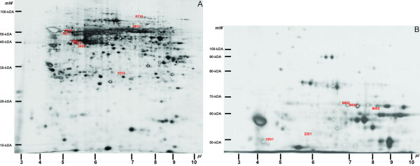
Methods: Here we describe a method to distinguish very likely endogenous from uncertain endogenous CK's in a mass spectrometry based proteomic study. In this study the CK identifications from 102 human corneal samples were compared with the number of human CK identifications found in 102 murine thymic lymphoma samples.
Results: It was anticipated that the CK's that were identified with a frequency of <5%, i.e. in less than one spot for every 20 spots analysed, are very likely to be endogenous and thereby represent a 'biologically significant' identification. CK's observed with a frequency >5% are uncertain endogenous since they may represent true endogenous CK's but the probability of contamination is high and therefore needs careful consideration. This was confirmed by comparison with a study of mouse samples where all identified human CK's are contaminants.
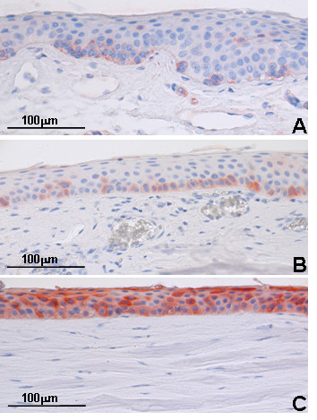
Conclusions: CK's 3, 4, 7, 8, 11, 12, 13, 15, 17, 18, 19, 20 and 23 are very likely to be endogenous proteins if identified in a corneal study, whilst CK's 1, 2e, 5, 6A, 9, 10, 14 and 16 may be endogenous although some are likely to be contaminants in a proteomic study. Further immunohistochemical analysis and a search of the current literature largely supported the distinction.
Figures
Similar articles
-
Immunoprecipitation and electrophoresis used to demonstrate and evaluate interference by CK-BB and atypical-CK's with CK-MB determinations by immunoinhibition.Clin Chem. 1984 Jan;30(1):167-8. Clin Chem. 1984. PMID: 6360419 No abstract available.
-
Coupling of the enzymic activities of myosin ATPase and creatine kinase and its role in muscular contraction.Indian J Biochem Biophys. 1989 Jun;26(3):148-52. Indian J Biochem Biophys. 1989. PMID: 2559887
-
Cytokeratin 8/18 expression indicates a poor prognosis in squamous cell carcinomas of the oral cavity.BMC Cancer. 2006 Jan 13;6:10. doi: 10.1186/1471-2407-6-10. BMC Cancer. 2006. PMID: 16412231 Free PMC article.
-
The creatine kinase response to resistance exercise.J Musculoskelet Neuronal Interact. 2014 Mar;14(1):68-77. J Musculoskelet Neuronal Interact. 2014. PMID: 24583542 Review.
-
Keratin expression by corneal and limbal stem cells during development.Exp Eye Res. 2020 Nov;200:108206. doi: 10.1016/j.exer.2020.108206. Epub 2020 Aug 31. Exp Eye Res. 2020. PMID: 32882212 Review.
Cited by
-
The Human Eye Proteome Project: perspectives on an emerging proteome.Proteomics. 2013 Aug;13(16):2500-11. doi: 10.1002/pmic.201300075. Proteomics. 2013. PMID: 23749747 Free PMC article. Review.
-
In-depth mass spectrometric mapping of the human vitreous proteome.Proteome Sci. 2013 May 20;11(1):22. doi: 10.1186/1477-5956-11-22. Proteome Sci. 2013. PMID: 23688336 Free PMC article.
-
Proteomic profile of vitreous in patients with tubercular uveitis.Tuberculosis (Edinb). 2021 Jan;126:102036. doi: 10.1016/j.tube.2020.102036. Epub 2020 Dec 3. Tuberculosis (Edinb). 2021. PMID: 33359883 Free PMC article.
-
A Critical Review of Bottom-Up Proteomics: The Good, the Bad, and the Future of this Field.Proteomes. 2020 Jul 6;8(3):14. doi: 10.3390/proteomes8030014. Proteomes. 2020. PMID: 32640657 Free PMC article. Review.
-
Human cornea proteome: identification and quantitation of the proteins of the three main layers including epithelium, stroma, and endothelium.J Proteome Res. 2012 Aug 3;11(8):4231-9. doi: 10.1021/pr300358k. Epub 2012 Jul 10. J Proteome Res. 2012. PMID: 22698189 Free PMC article.
References
-
- Uusitalo M, Kivela T. Development of cytoskeleton in neuroectodermally derived epithelial and muscle cells of the human eye. Invest Ophthalmol Vis Sci. 1995;36:2584–2591. - PubMed
Publication types
MeSH terms
Substances
LinkOut - more resources
Full Text Sources
Research Materials
