. 2011 Feb;121(2):658-70.
doi: 10.1172/JCI42974. Epub 2011 Jan 25.

Liver X receptor (LXR) mediates negative regulation of mouse and human Th17 differentiation

Affiliations

Liver X receptor (LXR) mediates negative regulation of mouse and human Th17 differentiation

Guoliang Cui et al. J Clin Invest. 2011 Feb.

Abstract

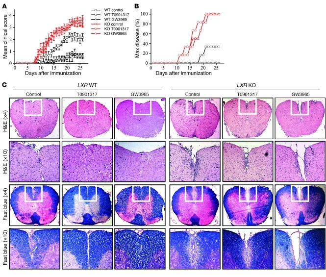
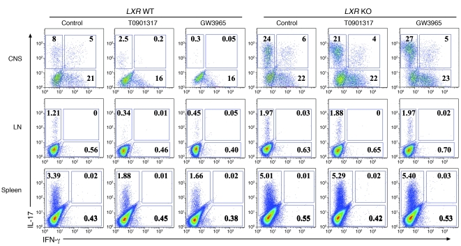
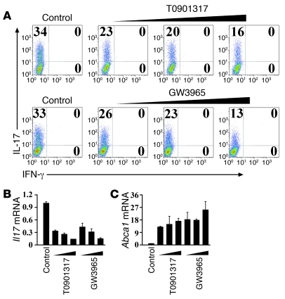
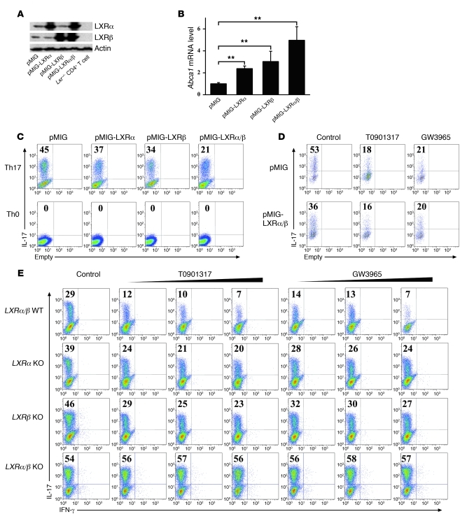
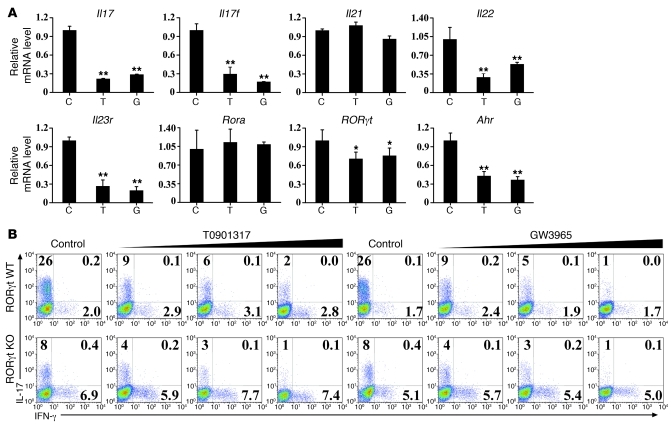
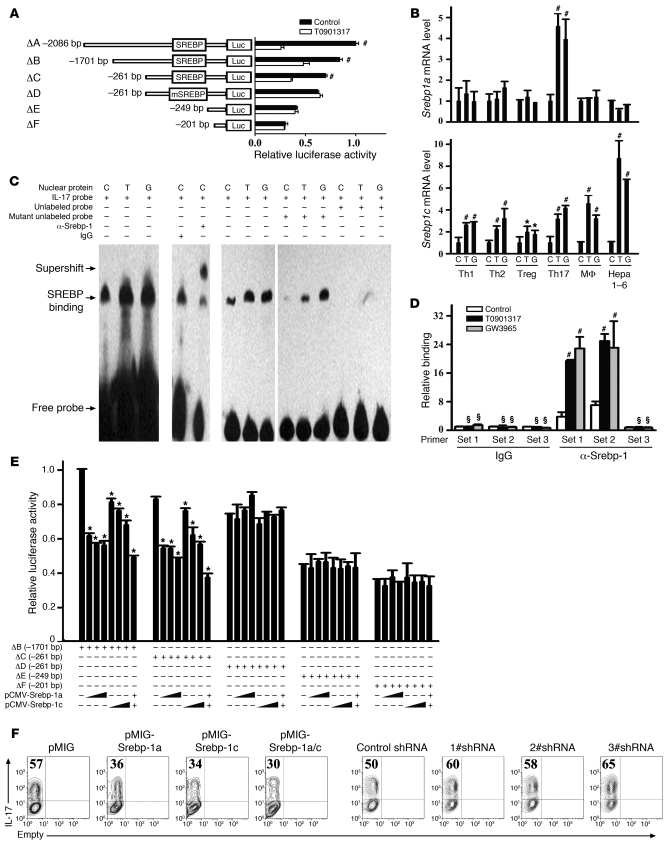
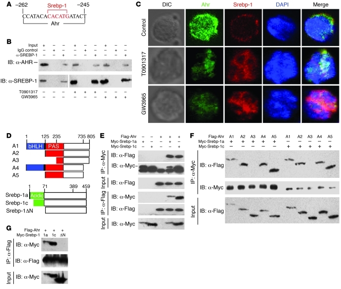
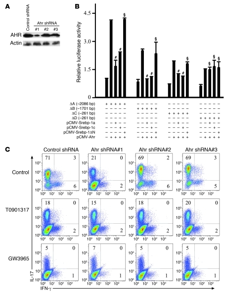
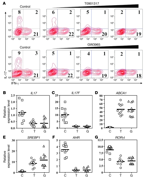
Th17 cells are a subset of CD4+ T cells with an important role in clearing certain bacterial and fungal pathogens. However, they have also been implicated in autoimmune diseases such as multiple sclerosis. Exposure of naive CD4+ T cells to IL-6 and TGF-β leads to Th17 cell differentiation through a process in which many proteins have been implicated. We report here that ectopic expression of liver X receptor (LXR) inhibits Th17 polarization of mouse CD4+ T cells, while LXR deficiency promotes Th17 differentiation in vitro. LXR activation in mice ameliorated disease in the experimental autoimmune encephalomyelitis (EAE) model of multiple sclerosis, whereas LXR deficiency exacerbated disease. Further analysis revealed that Srebp-1, which is encoded by an LXR target gene, mediated the suppression of Th17 differentiation by binding to the E-box element on the Il17 promoter, physically interacting with aryl hydrocarbon receptor (Ahr) and inhibiting Ahr-controlled Il17 transcription. The putative active site (PAS) domain of Ahr and the N-terminal acidic region of Srebp-1 were essential for this interaction. Additional analyses suggested that similar LXR-dependent mechanisms were operational during human Th17 differentiation in vitro. This study reports what we believe to be a novel signaling pathway underlying LXR-mediated regulation of Th17 cell differentiation and autoimmunity.


Figures

Figure 1
Figure 1. Oral administration of LXR agonists ameliorates EAE in LXR WT mice but not in LXR KO mice.
(A) EAE was induced in LXR KO mice or WT littermate control mice with or without daily oral administration of LXR agonist from day 3 after immunization to day 17 (6 or 7 mice per group). Data are expressed as mean ± SEM and represent 2 independent experiments with similar results. P = 0.009, WT control versus WT T0901317; P = 0.006, WT control group versus WT GW3965; P = 0.832, KO control versus KO T0901317; P = 0.651, KO control versus KO GW3965; P = 0.039, WT control versus KO control. (B) Maximum (Max) disease was set with a clinical score ≥3. P = 0.002, WT control versus WT T0901317; P = 0.002, WT control versus WT GW3965; P = 0.857, KO control versus KO T0901317; P = 0.807, KO control versus KO GW3965; P = 0.007, WT control versus KO control. (C) Histological staining of spinal cord sections from each group on day 15 after immunization. The white boxes in the upper panels (×4 objective) are enlarged in the lower panels (×10 objective). Total original magnification is ×40 and ×100, respectively.
Figure 2
Figure 2. Oral administration of LXR agonists decreases IL-17 production in vivo in LXR WT mice but not in LXR KO mice.
Intracellular staining of IL-17 and IFN-γ on CD4+ T cells isolated from the CNS, groin lymph nodes, and spleen, as indicated, 15 days after immunization (6 mice per group). Values in the quadrants indicate the percentage of IL-17+IFN-γ cells, IL-17+IFN-γ+ cells, and IL-17IFN-γ+ cells.
Figure 3
Figure 3. LXR activation suppresses in vitro Th17 cell differentiation.
(A) Mouse naive CD4+ T cells were cultured under Th17-inducing conditions for 4 days in the presence of LXR agonists T0901317 (0.5 μM, 1 μM, 2 μM) and GW3965 (2.5 μM, 5 μM, 10 μM) and subjected to intracellular staining of IL-17 and IFN-γ. This experiment was repeated more than 10 times with similar results. Values in the quadrants indicate the percentage of IL-17+IFN-γ cells, IL-17+IFN-γ+ cells, and IL-17IFN-γ+ cells. Cells from A were harvested for real-time PCR analysis of the Il17 (B) and Abca1 (C) mRNA levels. Data are expressed as mean ± SD in B and C.
Figure 4
Figure 4. LXRα/β negatively regulates in vitro Th17 differentiation.
(A) Mouse naive CD4+ T cells were cultured under Th17-inducing conditions with or without retroviral expression of LXRα and LXRβ for 4 days before protein isolation and Western blot analysis. Protein prepared from LXR KO CD4+ T cells was loaded as a control. (B) Cells from A were harvested for real-time PCR analysis of the Abca1 mRNA levels. **P < 0.01. Data in B are expressed as mean ± SD. (C) Naive CD4+ T cells were cultured under Th17- or Th0-inducing conditions with or without retroviral expression of LXRα and LXRβ for 4 days before IL-17 staining, with the gate set on GFP+CD4+ cells. This experiment was repeated at least 3 times with similar results. (D) Naive CD4+ T cells were cultured under Th17-inducing conditions with or without retroviral expression of LXRα/β and subjected to IL-17 staining with the gate set on GFP+CD4+ cells. LXR agonists T0901317 (2 μM) and GW3965 (10 μM) were added as indicated. This experiment was repeated at least 3 times with similar results. (E) Naive CD4+ T cells from LXR WT littermate control mice, LXRα KO, LXRβ KO, and LXRα/β KO mice were cultured under Th17-inducing conditions for 4 days in the presence of LXR agonists T0901317 (0.5 μM, 1 μM, 2 μM) and GW3965 (2.5 μM, 5 μM, 10 μM) before IL-17 and IFN-γ staining (3 mice per group). Values in CE indicate the percentage of IL-17+ cells.
Figure 5
Figure 5. LXR agonists suppress expression of RORγt and other Th17-related genes.
(A) Naive CD4+ T cells were cultured under Th17-inducing conditions for 4 days in the presence of vehicle control (C), T0901317 (T), and GW3965 (G) before cells were harvested for real-time PCR analysis of the genes as indicated above. Data are expressed as mean ± SD, and this experiment was repeated at least 3 times with similar results. *P < 0.05; **P < 0.01. (B) Naive CD4+ T cells isolated from RORγt WT mice or RORγt KO mice were cultured under Th17-inducing conditions with or without LXR agonists T0901317 (0 μM, 0.5 μM, 1 μM, 2 μM) and GW3965 (0 μM, 2.5 μM, 5 μM, 10 μM) for 4 days and subjected to intracellular staining of IL-17 and IFN-γ. This experiment was repeated at least 3 times with similar results. Values in the quadrants indicate the percentage of IL-17+IFN-γ cells, IL-17+IFN-γ+ cells, and IL-17IFN-γ+ cells.
Figure 6
Figure 6. LXR activation suppresses IL-17 transcriptional activity through the promotion of Srebp-1 binding to the E-box element on the Il17 promoter.
(A) Jurkat cells were transfected with fragments of the mouse Il17 promoter linked to a firefly luciferase construct and cultured with or without T0901317 (2 μM) before luciferase reporter assays. (B) Mouse naive CD4+ T cells were cultured under Th1-, Th2-, Treg-, or Th17-inducing conditions for 4 days in the presence of the indicated drugs before real-time PCR analysis of Srebp1a and Srebp1c. Mouse macrophage cell line RAW264.7 (MF) and hepatocyte cell line Hepa 1-6 were included as controls. (C and D) Srebp-1 binding to the E-box element on the Il17 promoter in in vitro differentiated Th17 cells was assessed by EMSA (C) and ChIP (D). The primers used to detect ChIP signals are schematically represented in Supplemental Figure 1. Statistical analysis was performed between the LXR agonist–treated and untreated groups. (E) Srebp-1a and Srebp-1c expression plasmids were transfected into Jurkat cells for luciferase reporter assay. (F) Naive CD4+ T cells were cultured under Th17-inducing conditions with or without retroviral overexpression or knockdown of Srebp-1a/c before IL-17 staining. This experiment was repeated at least 3 times with similar results. Values indicate the percentage of IL-17+ cells.*P < 0.05, #P < 0.01, §P > 0.05. Data are expressed as mean ± SD in A, B, D, and E.
Figure 7
Figure 7. Srebp-1 physically interacts with Ahr.
(A) Schematic representation of a fragment of the mouse Il17 promoter containing putative binding sites for Srebp-1 and Ahr. (B to C) Naive CD4+ T cells were cultured under Th17-inducing conditions with the indicated drugs before (B) coimmunoprecipitation and (C) confocal microscopy (×40 oil objective). A differential interference contrast (DIC) photo reveals the overall cellular structure, and the nucleus was stained with DAPI. The vertical line in B indicates that the lanes were run on the same gel but were noncontiguous. (D) Schematic representations of Ahr or Srebp-1 or deletion constructs (A1, A2, A3, A4, and A5 for Ahr; Srebp-1a, Srebp-1c, and Srebp-1ΔN for Srebp-1). bHLH, basic helix-loop-helix. (EG) Flag-tagged Ahr and Myc-tagged Srebp-1 (E), Flag-tagged Ahr or deletions and Myc-tagged Srebp-1 (F), or Flag-tagged Ahr and Myc-tagged Srebp-1 or deletion mutants (G) were transiently transfected into HEK293T cells for coimmunoprecipitation.
Figure 8
Figure 8. Srebp-1 interferes with Ahr-promoted ILI7 transcription.
(A) Western blot was performed using cells (C) to assess the efficiency of Ahr knockdown. (B) Ahr, Srebp-1a, or Srebp-1c constructs were transfected into Jurkat cells for luciferase reporter assay. #P < 0.01, §P > 0.05 compared with the Ahr single transfection group. Data are expressed as mean ± SD. (C) Mouse naive CD4+ T cells were cultured under Th17-inducing conditions with the indicated drugs and retroviral shRNA for 4 days before intracellular staining. This experiment was repeated at least 3 times with similar results. Values in the quadrants indicate the percentage of IL-17+IFN-γ cells, IL-17+IFN-γ+ cells, and IL-17IFN-γ+ cells.
Figure 9
Figure 9. LXR activation suppresses in vitro human Th17 cell differentiation.
(A) Human naive CD4+ T cells isolated from the peripheral blood mononuclear cells of a healthy donor were cultured under Th17-inducing conditions for 4 days in the presence of LXR agonists T0901317 (0.5 μM, 1 μM, 2 μM) and GW3965 (2.5 μM, 5 μM, 10 μM) and subjected to the intracellular staining of IL-17 and IFN-γ. Fifteen individual specimens were studied, with similar results. Values in the quadrants indicate the percentage of IL-17+IFN-γ cells, IL-17+IFN-γ+ cells, and IL-17IFN-γ+ cells. (BE) Cells from A were harvested for real-time PCR analysis of the mRNA levels of IL17 (B), IL17F (C), ABCA1 (D), SREBP1 (E), AHR (F), and RORγt (G). The y-axis in BG represents the relative expression level of genes using arbitrary unit. T0901317 decreased the IL17 (P < 0.01), IL17F (P < 0.01), AHR (P < 0.01), and RORγt (P < 0.05) mRNA levels and increased ABCA1 (P < 0.01) and SREBP1 (P < 0.01). GW3965 suppressed the expression of IL17 (P < 0.01), IL17F (P < 0.01), AHR (P < 0.01), and RORγt (P < 0.05) and increased the mRNA level of ABCA1 (P < 0.01) and SREBP1 (P < 0.01).

Comment in

Similar articles

Cited by

References

    1. Harrington LE, et al. Interleukin 17-producing CD4+ effector T cells develop via a lineage distinct from the T helper type 1 and 2 lineages. Nat Immunol. 2005;6(11):1123–1132. doi: 10.1038/ni1254. - DOI - PubMed
    1. Park H, et al. A distinct lineage of CD4 T cells regulates tissue inflammation by producing interleukin 17. Nat Immunol. 2005;6(11):1133–1141. doi: 10.1038/ni1261. - DOI - PMC - PubMed
    1. Veldhoen M, Hocking RJ, Atkins CJ, Locksley RM, Stockinger B. TGFbeta in the context of an inflammatory cytokine milieu supports de novo differentiation of IL-17-producing T cells. Immunity. 2006;24(2):179–189. - PubMed
    1. Yang XO, et al. STAT3 regulates cytokine-mediated generation of inflammatory helper T cells. J Biol Chem. 2007;282(13):9358–9363. doi: 10.1074/jbc.C600321200. - DOI - PubMed
    1. Ivanov II, et al. The orphan nuclear receptor RORgammat directs the differentiation program of proinflammatory IL-17+ T helper cells. Cell. 2006;126(6):1121–1133. - PubMed

Publication types

MeSH terms