. 2011 Feb 1;10(3):538-48.
doi: 10.4161/cc.10.3.14758. Epub 2011 Feb 1.

Threonine 48 in the BIR domain of survivin is critical to its mitotic and anti-apoptotic activities and can be phosphorylated by CK2 in vitro

Affiliations

Threonine 48 in the BIR domain of survivin is critical to its mitotic and anti-apoptotic activities and can be phosphorylated by CK2 in vitro

Rachel M A Barrett et al. Cell Cycle. .

Abstract

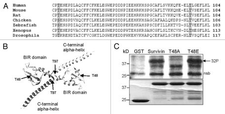
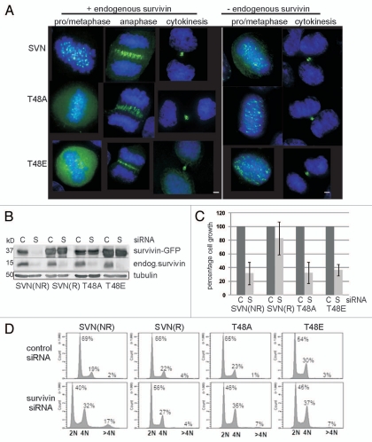
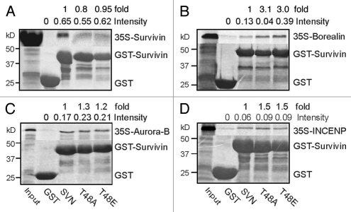
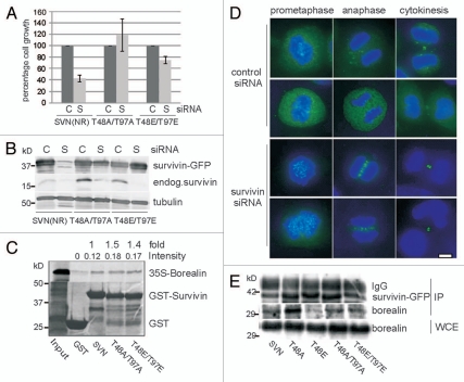
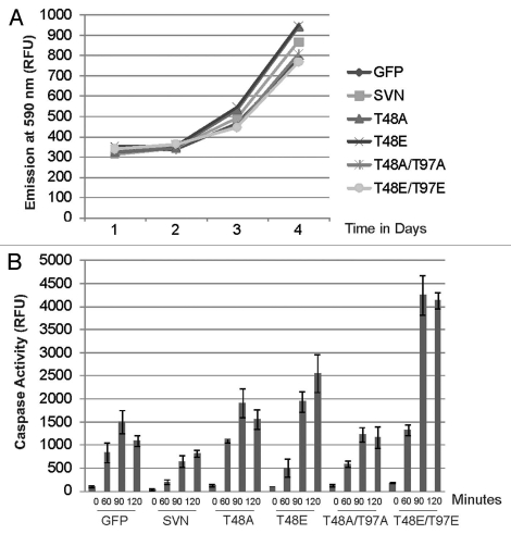
In this study we report that the protein kinase CK2 phosphorylates survivin specifically on threonine 48 (T48) within its BIR domain, and that T48 is critical to both the mitotic and anti-apoptotic roles of survivin. Interestingly, during mitosis T48 mutants localise normally, but are unable to support cell growth when endogenous survivin is removed by siRNA. In addition, while overexpression of survivin normally confers inhibition of TRAIL-mediated apoptosis, this protection is abolished by mutation of T48. Furthermore in interphase cells depletion of endogenous survivin causes redistribution of T48 mutants from the cytoplasm to the nucleus and treatment of cells expressing survivin-GFP with the CK2 inhibitor TBB phenocopies this nuclear redistribution. Finally, we show T48 mutants have increased affinity for borealin, and that this association and cell proliferation can be restored by introduction of a second mutation at T97. To our knowledge these data are the first to identify T48 as a key regulatory site on survivin, and CK2 as a mediator of its mitotic and anti-apoptotic functions.


Figures

Figure 1
Figure 1
Survivin is phosphorylated by CK2 on T48 in vitro. (A) Sequence alignment of residues 46 to 104 of human survivin with survivin from other species demonstrates conservation of T48 and T97 and their contextual residues. (B) Crystal structure of the survivin homodimer indicating the positions of T48 within the BIR domain, and T97 within the central linker. The molecular graphic was produced using the UCSF Chimera package (www.cgl.ucsf.edu/chimera). (C) In vitro kinase assay using GST, GST-survivin, GST-T48A and GST-T97A as potential substrates, incubated with recombinant CK2 in the presence of γ-ATP labelled 32P. Lower Coomassie Blue part indicates equality of loading. 32P radiolabel incorporated into survivin and T97A but not GST or T48A, demonstrating that T48 is the principle CK2 phosphotarget of survivin. Nsb denotes a non-specific band. Note that the nsb is of higher mw than GST and that GST itself is not phosphorylated.
Figure 2
Figure 2
Survivin CK2 mutants localise normally in mitosis but cannot support proliferation. (A) Exponentially growing HeLa cells expressing wild-type or mutant versions of survivin-GFP (green), as indicated, were fixed and stained to localise the DNA with DAPI (blue). All forms localized normally to the centromeres, midzone and midbody, during (pro)metaphase, anaphase and cytokinesis, when the endogenous protein was present (left parts). Upon removal of the endogenous form by RNAi, localization was also normal (right parts); however, no anaphase cells were observed. Bars 5 µm. (B) Endogenous survivin was depleted from the HeLa lines indicated using siRNA, and immunoblotted with anti-survivin antibodies 72 h post-depletion. C, control siRNA; S, survivin-specific siRNA. (NR indicates non-resistant to survivin siRNA; R indicates the resistant form). Both mutants bear resistance to survivin siRNA. (C) Survival of each cell line 72 h post-RNAi treatment was assessed by trypan blue exclusion. Cell growth of the survivin depleted sample is expressed as a percentage of the control cell number, which is given as 100%. The positive control (SVNR) restored cell growth to 80%, while lines expressing T48A and T48E showed comparable response to the negative control population (SVNNR), demonstrating that neither mutant could restore cell proliferation. Results are representative of three independent experiments with error bars indicating standard deviation. (D) FACS profiles of T48 mutants: Asynchronous cell lines were fixed and stained with propidium iodide 72 h post-siRNA. After depletion of the endogenous protein, both T48 mutants displayed a decrease in 2N cells (G1 phase), an increase in 4N cells (G2, M or binucleated), and a modest increase in cells with >4N (polyploid). FACS profiles are respresentative of two independent experiments.
Figure 3
Figure 3
TBB treatment phenocopies relocation of T48 mutants to the nucleus. (A–C) Interphase localization of GFP tagged SVN, T48A or T48E (green) in formaldehyde fixed, DAPI (blue) stained HeLa cell lines. All forms are distributed in the cytoplasm when endogenous survivin is present (left parts), but after its depletion with siRNA (right parts), T48A and T48E redistribute to the nucleus where they form punctuate foci. (D) Mitotic HeLa cells expressing survivin-GFP (green) were mock-treated (upper part), or treated with 100 µM TBB for 6 h to inhibit CK2 activity (lower part), then fixed and immunoprobed to localise the microtubules (red) and counterstained to localise the DNA (blue). Survivin-GFP localization during mitosis was unaltered by TBB. (E) Interphase cells expressing survivin-GFP were treated with TBB, which caused survivin-GFP to relocate to the nucleus and form discrete foci, some of which were centromeres, as indicated by CENP-A immunostaining (E, middle part inset, red). (E, right part) The localization of GFP alone is unaffected by TBB treatment. (F and G) EM9 cells expressing a TBB-sensitive CK2, or a TBB resistant version of CK2, V66A, were transfected with survivin-GFP then treated with 100 µM TBB for 48 h. Survivin-GFP redistributed from the cytoplasm to the nucleus in TBB-sensitive EM9 cells (F) but not in the TBB resistant cells (G). Bars 5 µm.
Figure 4
Figure 4
Mutation of T48 alters the affinity of survivin for borealin in vitro. Recombinant GST, GST-survivin, GST-T48A, or GST-T48E were bound to glutathione sepharose beads and incubated with the in vitro translated 35S-labelled CPP of interest: (A) survivin, (B) borealin, (C) aurora-B or (D) INCENP. While affinity for borealin was three-fold higher with the mutants (B), little difference was observed in their associations with survivin, aurora-B or INCENP. Data is representative of two independent experiments. Lane splicing was carried out in (A and D) to remove irrelevant samples. Pixel intensity of bands was measured using a Storm Phosphoimager, and normalized against the intensity of Coomassie GST-bands. Quantitation is presented as intensity in relative units, and fold difference from wild type.
Figure 5
Figure 5
Mutation of T97A/E alleviates repression of survivin activity conferred by T48A/E. (A and B) Cell lines indicated were subjected to RNAi with control (C) or survivin-specific (S) oligos for 72 h. (A) Number of viable cells is presented as a percentage of the control. (B) Immunoblot indicating the efficacy of depletion and confirming resistance of the double mutant forms. (C) Localization of double mutants (green) in the presence (upper parts) and absence (lower parts) of endogenous survivin in HeLa lines after fixation with formaldehyde and staining with DAPI (blue). Bar 5 µm. (D) GST-pull down with 35S-methionine labelled in vitro translated borealin with the GST-constructs indicated. (E) Asynchronous HeLa cells were cotransfected with pcDNA constructs encoding the relevant survivin-GFP variants and pcDNA-borealin. Anti-survivin antibodies and protein A/G beads were used to immunoprecipitate the ectopically expressed survivin variants and immunoblots interrogated for the presence of borealin. Exogenous borealin expression in the WCE, and IP'd GFP tagged version of survivin are shown to demonstrate equality in abundance.
Figure 6
Figure 6
T48 mutants and T48T97 double mutants abolish survivin's IAP activity. (A) A rezasurin cell proliferation assay was performed on cell lines overexpressing the survivin variants indicated; all cells grew normally. (B) Caspase-3 activity assay. Lysates prepared from the lines indicated after treatment for 0, 60, 90 or 120 minutes in TRAIL were incubated with Ac-DEVD-Amc, and fluorogenic release assessed using a Spectrophotometer in relative fluorescence units (RFU). Extracts prepared from cell lines expressing T48 mutants exhibited high caspase 3 activity, indicating that these forms were unable to protect cells against apoptosis. Second site mutations did not recover cytoprotection. Data is representative of three independent experiments. Average values are plotted with error bars indicating standard deviation within a single experiment performed in triplicate.

Comment in

Similar articles

Cited by

References

    1. Altieri DC. Survivin, cancer networks and pathway-directed drug discovery. Nat Rev Cancer. 2008;8:61–70. - PubMed
    1. Guha M, Altieri DC. Survivin as a global target of intrinsic tumor suppression. Cell Cycle. 2009;8:2708–2710. - PMC - PubMed
    1. Ruchaud S, Carmena M, Earnshaw WC. Chromosomal passengers: Conducting cell division. Nat Rev Mol Cell Biol. 2007;8:798–812. - PubMed
    1. Sun SC, Wei L, Li M, Lin SL, Xu BZ, Liang XW, et al. Perturbation of survivin expression affects chromosome alignment and spindle checkpoint in mouse oocyte meiotic maturation. Cell Cycle. 2009;8:3365–3372. - PubMed
    1. O'Connor DS, Grossman D, Plescia J, Li FZ, Zhang H, Villa A, et al. Regulation of apoptosis at cell division by p34(cdc2) phosphorylation of survivin. PNAS. 2000;97:13103–13107. - PMC - PubMed

Publication types

MeSH terms