. 2011 Mar 25;286(12):10814-24.
doi: 10.1074/jbc.M110.132514. Epub 2011 Jan 20.

Mutant A53T alpha-synuclein induces neuronal death by increasing mitochondrial autophagy

Affiliations

Mutant A53T alpha-synuclein induces neuronal death by increasing mitochondrial autophagy

Vinay Choubey et al. J Biol Chem. .

Abstract

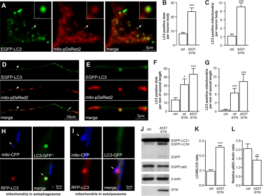
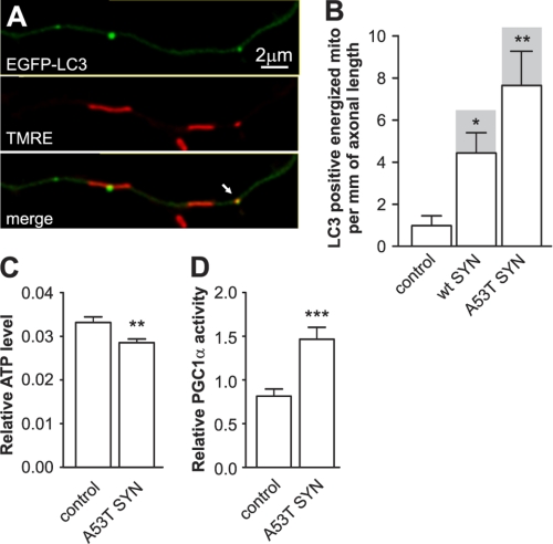
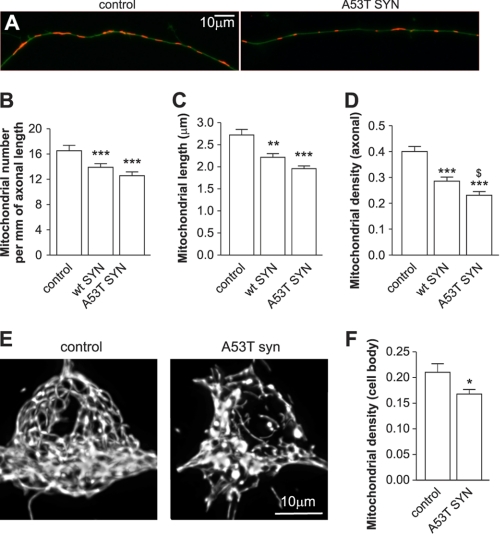
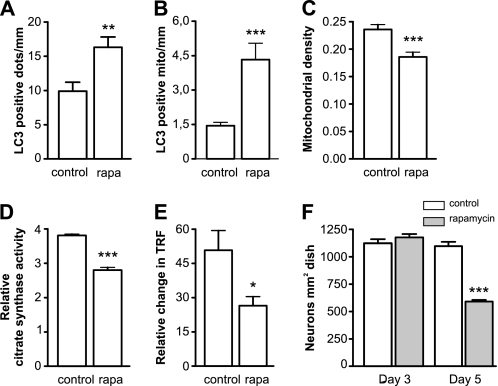
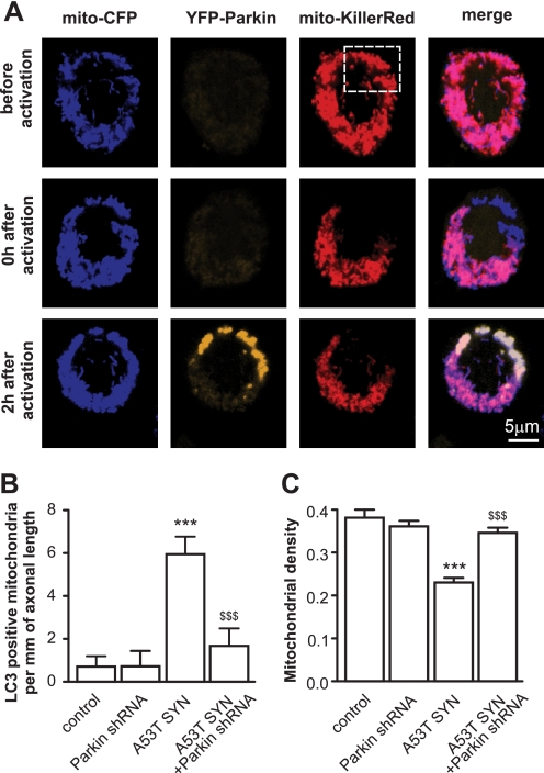
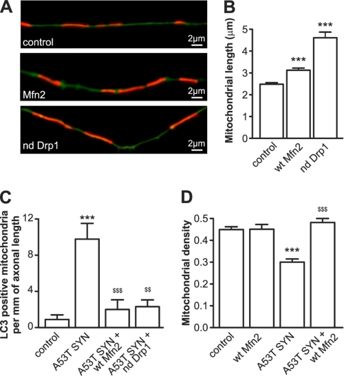
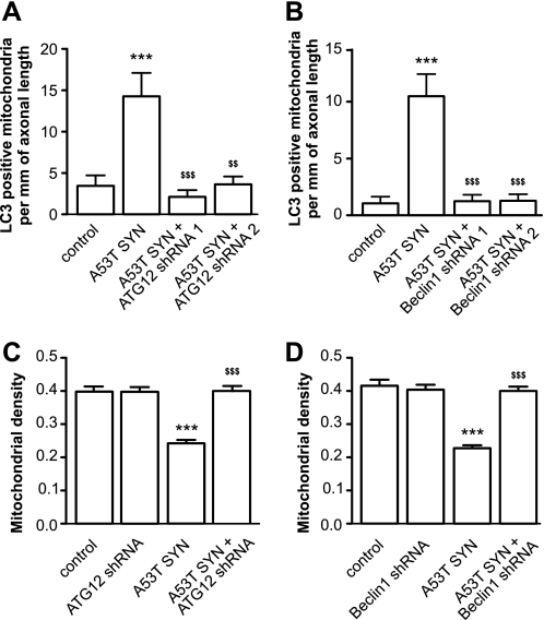
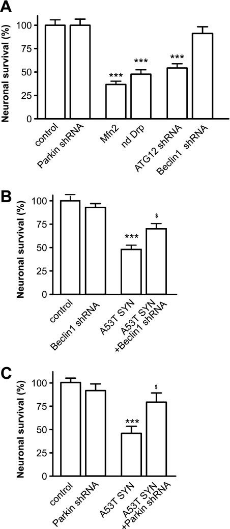
Parkinson disease is characterized by the accumulation of aggregated α-synuclein as the major component of the Lewy bodies. α-Synuclein accumulation in turn leads to compensatory effects that may include the up-regulation of autophagy. Another common feature of Parkinson disease (PD) is mitochondrial dysfunction. Here, we provide evidence that the overactivation of autophagy may be a link that connects the intracellular accumulation of α-synuclein with mitochondrial dysfunction. We found that the activation of macroautophagy in primary cortical neurons that overexpress mutant A53T α-synuclein leads to massive mitochondrial destruction and loss, which is associated with a bioenergetic deficit and neuronal degeneration. No mitochondrial removal or net loss was observed when we suppressed the targeting of mitochondria to autophagosomes by silencing Parkin, overexpressing wild-type Mitofusin 2 and dominant negative Dynamin-related protein 1 or blocking autophagy by silencing autophagy-related genes. The inhibition of targeting mitochondria to autophagosomes or autophagy was also partially protective against mutant A53T α-synuclein-induced neuronal cell death. These data suggest that overactivated mitochondrial removal could be one of the contributing factors that leads to the mitochondrial loss observed in PD models.


Figures

FIGURE 1.
FIGURE 1.
Overexpression of mutant A53T α-synuclein induces mitochondrial removal. Cortical neurons were transfected with plasmids expressing WT or mutant A53T α-synuclein, EGFP-LC3, and mito-pDsRed2 at day 2 after plating and visualized 6 days later. A, colocalization of EGFP-LC3 with mito-pDsRed2 in the cell body of neurons overexpressing mutant A53T α-synuclein. The left panel shows the EGFP-LC3 signal, the middle panel shows the mito-pDsRed2 signal, and the right panel shows the merged signals. The magnified inset shows the colocalization between the mitochondrial and autophagosome signal. Further analysis demonstrated an increased number of autophagosomes (B) as well as an increased number of mitochondria that colocalized with autophagosomes (C) per neuron body in neurons overexpressing mutant A53T α-synuclein overexpressing neurons (Mann-Whitney test). D, upper part of the panel shows the EGFP-LC3 signal, the middle panel shows the mito-pDsRed2 signal, and the lower panel shows the merged signals in neurites of neurons overexpressing mutant A53T α-synuclein. A magnified portion of the picture (E) demonstrates the exact colocalization between the mitochondrial and autophagosome signals. F and G, an increased number of autophagosomes (F) as well as increased number of mitochondria that colocalize with autophagosomes (G) per mm of axonal length in both the WT and mutant A53T α-synuclein overexpressing neurons (Kruskal-Wallis test followed by Dunn's test). H and I, neurons were transfected with A53T α-synuclein, mito-CFP, and RFP-LC3-GFP. H, Mito-CFP and RFP-LC3-GFP colocalize in the autophagosome while in the autolysosome (I) the GFP signal had faded in the acidic environment. J–L, overexpression of A53T α-synuclein enhances LC3-I conversion into LC3-II and decreases the levels of p62 (paired t test). *, p < 0.05; **, p < 0.01; ***, p < 0.001 versus control.
FIGURE 2.
FIGURE 2.
Overexpression of mutant A53T α-synuclein induces a bioenergetic deficit. Cortical neurons were transfected with plasmids expressing WT or mutant A53T α-synuclein and EGFP-LC3 and the mitochondria were stained with the mitochondrial membrane potential sensitive dye TMRE. A, colocalization of EGFP-LC3 (upper), TMRE (middle), and the superimposed channel (lower). B, colocalization analysis demonstrated a considerable increase of LC3-positive energized mitochondria in neurons overexpressing α-synucleins. Also note that the majority of LC3 positive mitochondria were energized (shaded columns show the total number of colocalizations; Kruskal-Wallis test followed by Dunn's test). C, neurons were cotransfected with plasmids expressing firefly luciferase and Renilla luciferase with or without mutant A53T α-synuclein at day 2 after plating. Firefly luciferase activity was measured in living neurons 24 h post-transfection using DMNPE-caged luciferin as a substrate followed by Renilla luciferase measurement (for normalization) in lysed neurons. The expression of mutant A53T α-synuclein decreased the ratio of firefly to Renilla luciferase activities, which indicated a decline in ATP levels. D, neurons were cotransfected with plasmids coding the Gal4-PGC-1α fusion protein, Gal4-UAS-luciferase reporter, and mutant A53T α-synuclein. Increased luciferase activity demonstrates that mutant A53T α-synuclein increases PGC-1α transcriptional activity (t test). *, p < 0.05; **, p < 0.01; ***, p < 0.001 versus control.
FIGURE 3.
FIGURE 3.
Overexpression of α-synuclein leads to mitochondrial loss. Cortical neurons were cotransfected with plasmids expressing WT or A53T mutated α-synuclein, GFP, and mitochondrially targeted pDsRed2 at day 2 after plating and visualized 6 days later. A, morphometric analysis of axonal mitochondria demonstrated that the mitochondrial number per mm axonal length (B), average mitochondrial length (C), and mitochondrial density (mitochondrial length per axonal length) (D) decreased in WT or mutant A53T α-synuclein overexpressing neurons. Note that the effect of mutant A53T α-synuclein is more pronounced than the WT protein (one way ANOVA followed by Newman Keuls test). To compare the mitochondrial density in the neuronal body between the control and mutant A53T α-synuclein expressing neurons, we performed a three-dimensional scan using a confocal microscope followed by three-dimensional reconstruction of the mitochondrial network in the neuronal body. E, three-dimensional reconstruction of the mitochondrial network from control neurons or mutant A53T α-synuclein expressing neurons. F, further stereological analysis of three-dimensional reconstructed mitochondrial networks in control neurons or in neurons expressing mutant A53T α-synuclein revealed a clear decrease in mitochondrial volume per cell volume (t test). *, p < 0.05; **, p < 0.01; and ***, p < 0.001 versus control; and $, p < 0.05 versus WT α-synuclein.
FIGURE 4.
FIGURE 4.
Rapamycin-induced mitochondrial removal and bioenergetic deficit. A and B, neurons were transfected with EGFP-LC3 and mitochondria-targeted pDsRed2 and treated with 100 nm rapamycin at day 2 after plating. Three days post-transfection, the LC3-positive dots were visualized. Rapamycin treatment led to a significant increase in LC3 positive dots (A) as well as LC3 positive mitochondria (B) per mm of axonal length (Mann Whitney test). C, neurons were transfected with GFP and mitochondria-targeted pDsRed2 and treated with rapamycin for 3 days, which led to a decrease in mitochondrial density. Rapamycin-treated neurons had decreased citrate synthase activity (D) and diminished oxygen consumption (E), both of which were normalized per number of intact neurons. Oxygen consumption was estimated by measuring the relative change in time resolved fluorescence (TRF) of the MitoXpressTM probe. F, rapamycin had no effect on neuronal survival after 3 days of treatment, but was clearly toxic after 5 days of treatment. Student's t test, *, p < 0.05; **, p < 0.01; and ***, p < 0.001 versus control.
FIGURE 5.
FIGURE 5.
The role of Parkin in mutant A53T α-synuclein-induced mitochondrial removal and mitochondrial loss. A, PC12 cells were transfected with plasmids expressing mito-CFP, YFP-Parkin, and mito-KillerRed. A subpopulation of mitochondria was then irradiated with 561 laser line, which caused a bleaching of the KillerRed signal. Two hours later, YFP-Parkin was localized to these mitochondria. For panels B and C, cortical neurons were transfected with plasmids expressing mutant A53T α-synuclein, EGFP or EGFP-LC3, mito-pDsRed2, and plasmids expressing shRNA against Parkin per the indicated combinations. B, suppression of Parkin inhibits mutant A53T α-synuclein-induced mitochondrial removal (Kruskal-Wallis test followed by Dunn's test). A plasmid encoding scrambled shRNA was used as a control for all of the shRNA experiments. C, suppression of Parkin also protects neurons from mutant A53T α-synuclein-induced mitochondrial loss (one way ANOVA followed by Newman Keuls test; interaction in all cases p < 0.0001, two way ANOVA). ***, p < 0.001 versus control, and $$, p < 0.01; $$$, p < 0.001 versus mutant A53T α-synuclein.
FIGURE 6.
FIGURE 6.
Inhibition of mitochondrial fragmentation protects against mutant A53T α-synuclein-induced mitochondrial loss. A–D, cortical neurons were transfected with plasmids expressing mutated A53T α-synuclein, mito-pDsRed2, EGFP (A, B, D) or EGFP-LC3 (C) and WT Mfn2 or a dominant negative Drp1. A and B, overexpression of WT Mfn2 and dominant negative Drp1 increase mitochondrial length. C, overexpression of WT Mfn2 or expression of dominant negative Drp1 inhibits mutant A53T α-synuclein-induced mitochondrial removal (Kruskal-Wallis test followed by Dunn's test; interaction in all cases p < 0.0001, two way ANOVA. D, overexpression of WT Mfn2 also protects neurons from mitochondrial loss induced by mutant A53T α-synuclein. ***, p < 0.001 versus control, and $$, p < 0.01; $$$, p < 0.001 versus A53T α-synuclein.
FIGURE 7.
FIGURE 7.
Inhibition of autophagy protects against mutant A53T α-synuclein-induced mitochondrial removal and mitochondrial loss. Cortical neurons were transfected with plasmids expressing mutant A53T α-synuclein, EGFP or EGFP-LC3, mito-pDsRed2, and plasmids expressing shRNA against Beclin1 or ATG12 as indicated. A and B, suppression of ATG12 and Beclin1 by specific shRNA decreases mutant A53T α-synuclein-induced mitochondrial removal. C and D, suppression of Beclin1 or ATG12 protects neurons from mutant A53T α-synuclein-induced mitochondrial loss (one way ANOVA followed by Newman Keuls test; interaction in all cases p < 0.0001, two way ANOVA). ***, p < 0.001 versus control and $$, p < 0.01; $$$, p < 0.001 versus mutant A53T α-synuclein.
FIGURE 8.
FIGURE 8.
Suppression of Parkin and Beclin1 protects neurons from mutant α-synuclein-induced neuronal death. Cortical neurons were transfected with plasmids expressing mutant A53T α-synuclein, EGFP, mito-pDsRed2, and the plasmids of interest at day 2 after plating. A, overexpression of WT Mfn2, dominant negative Drp1, or the suppression of ATG12 decreases the number of EGFP-expressing, viable neurons 6 days post-transfection, while the suppression of Parkin or Beclin1 was relatively nontoxic. B and C, suppression of Beclin1 (B) and Parkin (C) partially protects against mutant A53T α-synuclein-induced neuronal loss. EGFP-positive viable neurons from the control at 6 days post-transfection were set as 100%. One way ANOVA followed by Newman Keuls test. **, p < 0.01; ***, p < 0.001 versus control; and $, p < 0.05; $$$, p < 0.001 versus mutant A53T α-synuclein.

Similar articles

Cited by

References

    1. Schapira A. H., Cooper J. M., Dexter D., Jenner P., Clark J. B., Marsden C. D. (1989) Lancet 1, 1269. - PubMed
    1. Henchcliffe C., Beal M. F. (2008) Nat. Clin. Pract. Neurol. 4, 600–609 - PubMed
    1. Ayala A., Venero J. L., Cano J., Machado A. (2007) Front Biosci. 12, 986–1007 - PubMed
    1. Schapira A. H. (2008) Lancet Neurol. 7, 97–109 - PubMed
    1. Anglade P., Vyas S., Javoy-Agid F., Herrero M. T., Michel P. P., Marquez J., Mouatt-Prigent A., Ruberg M., Hirsch E. C., Agid Y. (1997) Histol. Histopathol. 12, 25–31 - PubMed

Publication types

MeSH terms