. 2011 Feb;121(2):582-92.
doi: 10.1172/JCI44907. Epub 2011 Jan 18.

Maternal T cells limit engraftment after in utero hematopoietic cell transplantation in mice

Affiliations

Maternal T cells limit engraftment after in utero hematopoietic cell transplantation in mice

Amar Nijagal et al. J Clin Invest. 2011 Feb.

Abstract

Transplantation of allogeneic stem cells into the early gestational fetus, a treatment termed in utero hematopoietic cell transplantation (IUHCTx), could potentially overcome the limitations of bone marrow transplants, including graft rejection and the chronic immunosuppression required to prevent rejection. However, clinical use of IUHCTx has been hampered by poor engraftment, possibly due to a host immune response against the graft. Since the fetal immune system is relatively immature, we hypothesized that maternal cells trafficking into the fetus may pose the true barrier to effective IUHCTx. Here, we have demonstrated that there is macrochimerism of maternal leukocytes in the blood of unmanipulated mouse fetuses, with substantial increases in T cell trafficking after IUHCTx. To determine the contribution of these maternal lymphocytes to rejection after IUHCTx, we bred T and/or B cell-deficient mothers to wild-type fathers and performed allogeneic IUHCTx into the immunocompetent fetuses. There was a marked improvement in engraftment if the mother lacked T cells but not B cells, indicating that maternal T cells are the main barrier to engraftment. Furthermore, when the graft was matched to the mother, there was no difference in engraftment between syngeneic and allogeneic fetal recipients. Our study suggests that the clinical success of IUHCTx may be improved by transplanting cells matched to the mother.


Figures

Figure 1
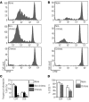
Figure 1. Lineage analysis of donor- and host-derived leukocytes in congenic recipients.
Multilineage engraftment of FL-derived hematopoietic cells was seen in primary (thymus and BM) and secondary (spleen and lymph node [LN]) lymphoid organs and peripheral blood at 43–57 weeks after in utero transplantation (n ≥ 5 mice per group). Donor- and host-derived double-positive (DP: CD4+CD8+) and single-positive (SP: CD4+CD8 or CD4CD8+) thymocytes, T lymphocytes (T, CD4, and CD8), B lymphocytes (B), and granulocytes (Gr) are shown as a percentage of their respective CD45+ leukocyte gate (donor, CD45.1; host, B6; CD45.2). No significant differences were observed between the percentages of donor and host leukocyte subpopulations, with the exception of a decreased percentage of donor-derived B cells in spleen. *P < 0.05 by t test.
Figure 2
Figure 2. The adaptive immune response limits engraftment after allogeneic IUHCTx.
(A) Frequency of chimerism (number of chimeric animals/number of surviving animals) after in utero transplantation of B6 FL cells into congenic (CD45.1 into B6 [CD45.1/B6], n = 17), allogeneic (B6 into BALB/c [B6/BALB/c], n = 43), or immunodeficient (B6 into BABL/c.Rag1–/– [B6/Rag1–/–], n = 9) recipients. *P < 0.005, c2 test for allogeneic versus congenic. (B) Levels of chimerism in individual engrafted animals at 4 weeks after in utero transplantation (CD45.1/B6, n = 18; B6/BALB/c, n = 16; B6/Rag1–/–, n = 10). **P < 0.05, ANOVA with Tukey’s multiple comparison test. (C) Change in levels of chimerism over time when normalized to the initial level of chimerism at 4 weeks after transplantation (CD45.1/B6, n = 18; B6/BALB/c, n = 10; B6/Rag1–/–, n = 10). *P < 0.05 comparing CD45.1/B6 and B6/BALB/c, and CD45.1/B6 and B6/Rag1–/– using ANOVA with Tukey’s multiple comparison test.
Figure 3
Figure 3. Chimeras are tolerant to donor alloantigen.
Lymphocytes harvested from spleens and lymph nodes of naive mice, injected non-chimeras, and chimeras were labeled with CFSE and injected into (AC) B6 × BALB/c or (D) C3H × DBA/2 (third-party control) F1 recipients. Representative flow cytometric histograms showing CFSE profiles after gating on donor-derived (A) CD4+ or (B) CD8+ T lymphocytes are shown. (C) Percentage of alloreactive T cells in naive mice, non-chimeras, and chimeras. The data shown are representative of at least 4 independent experiments (naive, n = 10; non-chimera, n = 7; chimera, n = 10). *P < 0.05 comparing chimeras with naive mice and non-chimeras using ANOVA with Tukey’s multiple comparison test. (D) Percentage of donor-derived proliferating cells (%CFSElow) from naive and chimeric mice in response to a third-party antigen. Data are representative of at least 2 independent experiments (naive, n = 3; chimera, n = 5).
Figure 4
Figure 4. Maternal macrochimerism in mid-gestation fetal blood.
(A) The breeding scheme used to identify maternal leukocytes in fetal blood. (B) Representative flow cytometric plots depicting the profile of CD45.2+ (maternal, left panel), CD45.1+/CD45.2+ (adult control, middle panel), and E15.5 fetal blood (right panel; F, fetal; M, maternal). Lineage analysis of maternal leukocytes found in fetal blood (gate M in B) was performed using cell surface markers for (C) innate and (D) adaptive immune cells. (C) The gating strategy for identifying innate immune cells involved first detecting Gr-1+ or F4/80+ leukocytes. NK cells were identified among the Gr-1F4/80 cells. Gr1F4/80NK1.1 cells were further divided into CD11c+ and B220+ leukocytes. (D) Adaptive immune cells were characterized by identifying CD3+ and CD19+ maternal leukocytes. The CD3+ subpopulation was further characterized based on CD4 and CD8 expression. (E) Percentages of various leukocyte subsets found in the mother (maternal) and in the fetus (trafficked) at E12.5–E15.5 (n ≥ 3; *P < 0.01, **P < 1 × 10⁻⁸ by t test). (F) Percentage of maternal leukocytes (number of CD45.2+ cells/total CD45+ cells) in fetal circulation at various embryonic days of gestation (E12.5, n = 1; E13.5, n = 4; E14.5, n = 8; E15.5, n = 5; E18.5, n = 14; E20, n = 12; E22, n = 3). There was a significant negative correlation between maternal macrochimerism and gestational age (Pearson r = –0.94, P = 0.002).
Figure 5
Figure 5. Maternal-fetal cellular trafficking after fetal intervention.
B6 mothers were mated with CD45.1 fathers, and the CD45.1+/CD45.2+ fetuses were injected with allogeneic NOD.CD45.1 FL cells or PBS on E14.5. Injected (and uninjected control) fetuses were sacrificed on E18.5–E19.5, and the number of maternal leukocytes (CD45.2+) in fetal blood was quantified. (A) Flow cytometric analysis of donor (gate D), maternal (gate M), and fetal (gate F) leukocytes. (B) Frequency of fetuses with circulating maternal leukocytes (number of fetuses with circulating maternal leukocytes/total number of fetuses) after PBS injection (n = 11/33, 33%) and allogeneic FL injection (n = 12/21, 57%) and in age-matched uninjected controls (n = 0/21, 0%). (C) Percentage of maternal leukocytes (CD45.2+ maternal leukocytes/total CD45.2+ cells) in fetal circulation after PBS (n = 11) and allogeneic FL injection (n = 12). (D) Lineage analysis of trafficking maternal cells shown as percentage of maternal leukocytes (e.g., trafficked maternal Gr-1+ cells/total trafficked maternal leukocytes). *P < 0.005, **P < 0.0001 by t test. (E) Lineage analysis of trafficking maternal cells shown as the percentage of maternal cells contributing to each of the leukocyte subsets in fetal circulation (e.g., trafficked maternal Gr-1+ cells/total number of fetal and trafficked maternal Gr-1+ cells). *P < 0.05 by t test.
Figure 6
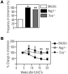
Figure 6. The rejection of in utero transplanted allogeneic hematopoietic cells occurs independent of maternal B cells and maternal alloantibodies.
(A) Frequency of chimerism after IUHCTx of B6 FL cells into fetuses born to a wild-type BALB/c father and either a wild-type BALB/c mother (n = 43) or a B cell–deficient (JHD) mother (n = 9). (B) Change in levels of chimerism over time in engrafted animals when normalized to the initial level of chimerism at 4 weeks after transplantation (BALB/c mother, n = 10; JHD mother, n = 4). (C) Frequency of chimerism in pups fostered by naive mothers (BALB/c fostered, n = 20) and in non-fostered pups (BALB/c, n = 43) after IUHCTx with B6 FL. (D) Serum from BALB/c mothers whose fetuses received allogeneic IUHCTx (n ≥ 10) was analyzed by flow cytometry to quantify total serum IgM (left panels) and IgG (right panels) alloantibody. Comparison groups include naive (n = 7) and sensitized (Sens., n ≥ 3) mice. (E) Total IgM (left panel) and IgG (right panel) alloantibody production at 1, 2, 4, and 6 weeks after sensitization is shown as the MFI relative to a no-serum sample (relative MFI). *P < 0.05 comparing IUHCTx with naive by ANOVA with Tukey’s multiple comparison test.
Figure 7
Figure 7. Maternal T cells limit engraftment and contribute to ongoing losses in chimerism after allogeneic IUHCTx.
Engraftment after transplantation of B6 FL cells into fetuses born to wild-type BALB/c fathers and either wild-type BALB/c or immunodeficient (Rag1–/– or Tcra–/–) mothers. (A) Frequency of chimerism (BALB/c, n = 43; Rag1–/–, n = 10; Tcra–/–, n = 21). *P < 0.005 comparing Rag1–/– with BALB/c, c2 test; **P < 0.001 comparing Tcra–/– with BALB/c, c2 test. (B) Change in levels of chimerism over time when normalized to the initial level of chimerism at 4 weeks after transplantation (BALB/c, n = 10; Rag1–/–, n = 12; Tcra–/–, n = 13). *P < 0.05 comparing BALB/c and Tcra–/–; **P < 0.05 comparing both BALB/c and Tcra–/–, and BALB/c and Rag1–/–. Comparisons were performed using ANOVA with Tukey’s multiple comparison test.
Figure 8
Figure 8. MHC matching between mother and graft improves engraftment in MHC-mismatched fetuses.
(A) Breeding scheme used to MHC match the mother with donor cells. B6 × BALB/c F1 (H-2b/d) female mice were mated to BALB/c (H-2d/d) males, and fetuses received B6 H-2b/b FL cells. (B) Frequency of chimerism among fetuses that were MHC matched (H-2b/d, n = 11) or mismatched (H-2d/d, n = 12) to the donor graft.

Similar articles

Cited by

References

    1. Billingham RE, Brent L, Medawar PB. Actively acquired tolerance of foreign cells. Nature. 1953;172(4379):603–606. doi: 10.1038/172603a0. - DOI - PubMed
    1. Merianos D, Heaton T, Flake AW. In utero hematopoietic stem cell transplantation: progress toward clinical application. Biol Blood Marrow Transplant. 2008;14(7):729–740. doi: 10.1016/j.bbmt.2008.02.012. - DOI - PubMed
    1. Kim HB, Shaaban AF, Yang EY, Liechty KW, Flake AW. Microchimerism and tolerance after in utero bone marrow transplantation in mice. J Surg Res. 1998;77(1):1–5. doi: 10.1006/jsre.1997.5255. - DOI - PubMed
    1. Hayashi S, Peranteau WH, Shaaban AF, Flake AW. Complete allogeneic hematopoietic chimerism achieved by a combined strategy of in utero hematopoietic stem cell transplantation and postnatal donor lymphocyte infusion. Blood. 2002;100(3):804–812. - PubMed
    1. Hayashi S, et al. Mixed chimerism following in utero hematopoietic stem cell transplantation in murine models of hemoglobinopathy. Exp Hematol. 2003;31(2):176–184. doi: 10.1016/S0301-472X(02)01024-X. - DOI - PubMed

Publication types