

Dishevelled interacts with the DIX domain polymerization interface of Axin to interfere with its function in down-regulating β-catenin
- PMID: 21245303
- PMCID: PMC3033301
- DOI: 10.1073/pnas.1017063108
Dishevelled interacts with the DIX domain polymerization interface of Axin to interfere with its function in down-regulating β-catenin
Abstract
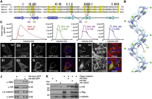
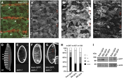
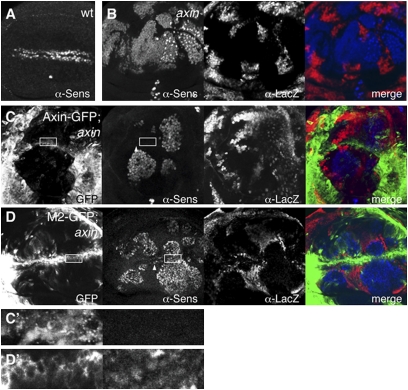
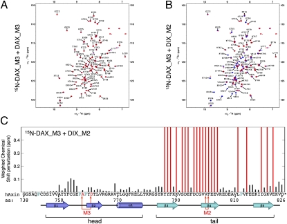
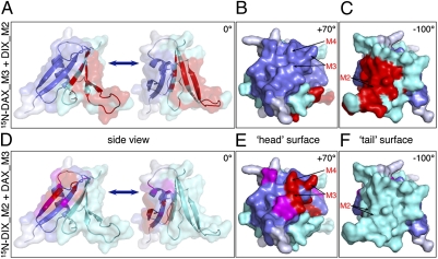
Wnt/β-catenin signaling controls numerous steps in normal animal development and can also cause cancer if inappropriately activated. In the absence of Wnt, β-catenin is targeted continuously for proteasomal degradation by the Axin destruction complex, whose activity is blocked upon Wnt stimulation by Dishevelled, which recruits Axin to the plasma membrane and assembles it into a signalosome. This key event during Wnt signal transduction depends on dynamic head-to-tail polymerization by the DIX domain of Dishevelled. Here, we use rescue assays in Drosophila tissues and functional assays in human cells to show that polymerization-blocking mutations in the DIX domain of Axin disable its effector function in down-regulating Armadillo/β-catenin and its response to Dishevelled during Wnt signaling. Intriguingly, NMR spectroscopy revealed that the purified DIX domains of the two proteins interact with each other directly through their polymerization interfaces, whereby the same residues mediate both homo- and heterotypic interactions. This result implies that Dishevelled has the potential to act as a "natural" dominant-negative, binding to the polymerization interface of Axin's DIX domain to interfere with its self-assembly, thereby blocking its effector function.
Conflict of interest statement
The authors declare no conflict of interest.
Figures
Similar articles
-
The Adenomatous polyposis coli tumour suppressor is essential for Axin complex assembly and function and opposes Axin's interaction with Dishevelled.Open Biol. 2011 Nov;1(3):110013. doi: 10.1098/rsob.110013. Open Biol. 2011. PMID: 22645652 Free PMC article.
-
A direct heterotypic interaction between the DIX domains of Dishevelled and Axin mediates signaling to β-catenin.Sci Signal. 2019 Dec 10;12(611):eaaw5505. doi: 10.1126/scisignal.aaw5505. Sci Signal. 2019. PMID: 31822591 Free PMC article.
-
Stability elements in the LRP6 cytoplasmic tail confer efficient signalling upon DIX-dependent polymerization.J Cell Sci. 2010 May 1;123(Pt 9):1588-99. doi: 10.1242/jcs.067546. Epub 2010 Apr 13. J Cell Sci. 2010. PMID: 20388731 Free PMC article.
-
The cellular story of dishevelleds.Croat Med J. 2014 Oct;55(5):459-67. doi: 10.3325/cmj.2014.55.459. Croat Med J. 2014. PMID: 25358879 Free PMC article. Review.
-
New steps in the Wnt/beta-catenin signal transduction pathway.Recent Prog Horm Res. 2000;55:225-36. Recent Prog Horm Res. 2000. PMID: 11036939 Review.
Cited by
-
The Wnt/β-catenin pathway in human fibrotic-like diseases and its eligibility as a therapeutic target.Mol Cell Ther. 2015 Jan 30;3:1. doi: 10.1186/s40591-015-0038-2. eCollection 2015. Mol Cell Ther. 2015. PMID: 26056602 Free PMC article.
-
tRNA-derived fragment tRF-1020 ameliorates diabetes-induced retinal microvascular complications.J Cell Mol Med. 2022 Oct;26(20):5257-5266. doi: 10.1111/jcmm.17555. Epub 2022 Sep 20. J Cell Mol Med. 2022. PMID: 36128646 Free PMC article.
-
Exploring DIX-DIX Homo- and Hetero-Oligomers in Wnt Signaling with AlphaFold2.Cells. 2024 Oct 3;13(19):1646. doi: 10.3390/cells13191646. Cells. 2024. PMID: 39404409 Free PMC article.
-
WISP1 and Macrophage Migration Inhibitory Factor in Respiratory Inflammation: Novel Insights and Therapeutic Potentials for Asthma and COPD.Int J Mol Sci. 2024 Sep 18;25(18):10049. doi: 10.3390/ijms251810049. Int J Mol Sci. 2024. PMID: 39337534 Free PMC article. Review.
-
Tankyrase Sterile α Motif Domain Polymerization Is Required for Its Role in Wnt Signaling.Structure. 2016 Sep 6;24(9):1573-81. doi: 10.1016/j.str.2016.06.022. Epub 2016 Aug 4. Structure. 2016. PMID: 27499439 Free PMC article.
References
Publication types
MeSH terms
Substances
Grants and funding
LinkOut - more resources
Full Text Sources
Molecular Biology Databases
Research Materials
