. 2011 Jan 24;192(2):219-27.
doi: 10.1083/jcb.201008076. Epub 2011 Jan 17.

ATR-mediated phosphorylation of DNA polymerase η is needed for efficient recovery from UV damage

Affiliations

ATR-mediated phosphorylation of DNA polymerase η is needed for efficient recovery from UV damage

Thomas Göhler et al. J Cell Biol. .

Abstract

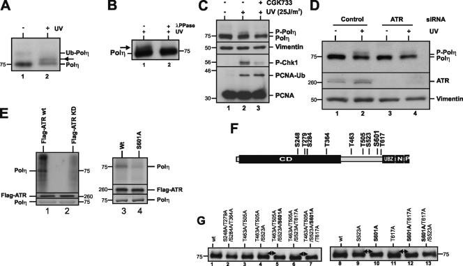
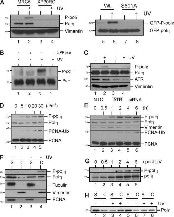
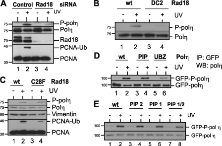
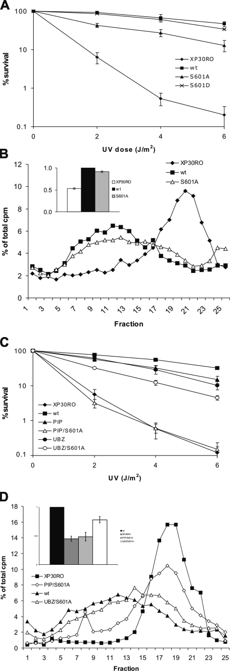
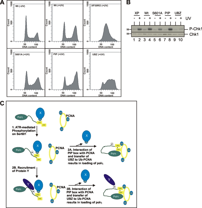
DNA polymerase η (polη) belongs to the Y-family of DNA polymerases and facilitates translesion synthesis past UV damage. We show that, after UV irradiation, polη becomes phosphorylated at Ser601 by the ataxia-telangiectasia mutated and Rad3-related (ATR) kinase. DNA damage-induced phosphorylation of polη depends on its physical interaction with Rad18 but is independent of PCNA monoubiquitination. It requires the ubiquitin-binding domain of polη but not its PCNA-interacting motif. ATR-dependent phosphorylation of polη is necessary to restore normal survival and postreplication repair after ultraviolet irradiation in xeroderma pigmentosum variant fibroblasts, and is involved in the checkpoint response to UV damage. Taken together, our results provide evidence for a link between DNA damage-induced checkpoint activation and translesion synthesis in mammalian cells.


Figures

Figure 1.
Figure 1.
Polη is phosphorylated at Ser601 after UV irradiation. (A) Anti-polη Western blot analysis of cell lysates from either unirradiated or UV-irradiated (25 J/m2) MRC5 cells, incubated for 6 h. The band of ubiquitinated polη (only seen in unirradiated cells) is indicated and the band of interest (only in irradiated cells) is denoted with an arrow. (B) polη was immunoprecipitated from UV-irradiated MRC5 cells (25 J/m2), incubated for 6 h, and immunoprecipitates were split in half and treated with or without λPPase. (C) MRC5 cells were treated with and without an ATM/ATR inhibitor (10 µM CGK733). Cells were UV irradiated (25 J/m2) and cell lysates were analyzed 6 h after irradiation. (D) MRC5 cells were transfected with control (lanes 1 and 2) or ATR siRNA (lanes 3 and 4), UV irradiated 48 h later (25 J/m2), and incubated for a further 6 h. (E) In vitro kinase assay with wild-type Flag-ATR (lanes 1, 3, 4) and a kinase-dead Flag-ATR (lane 2) immunoprecipitated from HEK 293 cells. The substrate was recombinant wild-type His6-polη (lanes 1–3) or S601A mutant (lane 4). Bottom panels show Western blot of immunoprecipitated wild-type and kinase-dead Flag-ATR and Coomassie staining of recombinant polη. (F) Schematic of polη. CD, catalytic domain; UBZ, ubiquitin-binding zinc-finger motif; N, nuclear localization sequence; P, PIP box PCNA-binding motif. Potential ATR phosphorylation sites (S/TQ) are indicated. (G) MRC5 cells were transfected with different polη mutant constructs. 24 h after transfection cells were UV irradiated (25 J/m2), and 6 h later cell lysates were prepared and analyzed. Double-headed arrows indicate loss of phosphorylated polη to the right and presence to the left.
Figure 2.
Figure 2.
Use of phospho-specific antibody to characterize polη phosphorylation. (A) Analysis of lysates from cells that were either unirradiated or UV irradiated (25 J/m2) and incubated for 6 h. MRC5 (lanes 1 and 2), XP30RO (lanes 3 and 4), or MRC5 cells transfected with eGFP-polη (lanes 5 and 6) or with eGFP-polη-S601A (lanes 7 and 8). (B) polη immunoprecipitates from irradiated (25 J/m2) and unirradiated MRC5 cells, incubated for 6 h, were either treated or untreated with λPPase. (C) MRC5 cells were depleted of ATR and treated as described in Fig. 1 D. (D) MRC5 cells were either unirradiated or UV irradiated and incubated for 6 h. (E) MRC5 cells either unirradiated or irradiated (20 J/m2) were incubated for the indicated times. (F) MRC5 cells either unirradiated or UV irradiated (25 J/m2) were incubated for 6 h and then extracted with Triton X-100. Triton-soluble fractions (S, lanes 1 and 3) and insoluble chromatin fractions (C, lanes 2 and 4) were subjected to Western blot analysis. Tubulin and vimentin were used as cytoplasmic and nuclear marker, respectively. (G) UV-irradiated cells were incubated for different times after UV irradiation and extracted with Triton X-100. Western blots of chromatin fractions were probed with antibody to the phosphorylated form and to total polη. (H) The experiment of Fig. 1 F was repeated but using cells expressing either wild-type polη (lanes 1–4) or S601A mutant (lanes 5–8) and blots probed with antibody to total polη.
Figure 3.
Figure 3.
Polη phosphorylation depends on interaction with Rad18. (A) MRC5 cells were incubated with control (lanes 1 and 2) or Rad18 siRNA (lanes 3 and 4). Cells were either unirradiated or UV irradiated (20 J/m2) and incubated for 6 h. (B) MRC5 cells were transfected with a wild-type Rad18 construct (lanes 1 and 2) or Rad18 DC2 (lanes 3 and 4) together with GFP as a transfection marker. Sorted cells were either unirradiated or UV irradiated and incubated for 6 h. (C) As in B, but using Rad18 C28F (lanes 3 and 4). (D) MRC5 cells were transfected with wild-type EGFP-polη (lanes 1 and 2), PIP box mutant (lanes 3 and 4), or UBZ mutant (lanes 5 and 6). Cells were either unirradiated or UV irradiated (20 J/m2), incubated for 6 h, and polη proteins were immunoprecipitated with a GFP antibody, followed by immunoblotting with anti-polη and the P-polη antibody. (E) As in D, but using cells expressing polη that was wild-type, mutated at PIP1 (aa 443-444), PIP2 (aa 707,708), or both.
Figure 4.
Figure 4.
Polη phosphorylation is necessary for efficient bypass of UV-induced photoproducts. (A) UV survival assay of XP30RO-derived cell lines expressing EGFP-polη, EGFP-polη-S601A, and EGFP-polη-S601D plated in the presence of 0.375 mM caffeine. Error bars denote SD of three experiments. (B) Alkaline sucrose sedimentation analysis of DNA from cells that were UV irradiated (8 J/m2), pulsed for 30 min with 3H-thymidine, and chased for 150 min, all in the presence of 0.375 mM caffeine. XP30RO expressing EGFP-polη-S601A (open triangles) was compared with one cell line proficient in damage bypass (XP30RO complemented with wild-type EGFP-polη, squares) and one defective (XP30RO, diamonds). The inset shows the average molecular weights of the distributions ± SD from three experiments. (C) UV survival assay of XP30RO-derived cell lines expressing the indicated polη mutant constructs. Error bars denote SD of three experiments. (D) As in B, but with cells expressing PIP/S601A and UBZ/S601A double mutants.
Figure 5.
Figure 5.
Polη phosphorylation, cell cycle progression, and checkpoint activation. (A) Cell cycle profiles of XP30RO cells complemented with EGFP-polη before irradiation (0 h) and of XP30RO cells complemented with the indicated wild-type or mutant forms of polη 24 h after UV irradiation (8 J/m2). (B) Unirradiated or UV-irradiated (15 J/m2) XP30RO-derived cell lines expressing EGFP-polη, either wild-type or indicated mutant forms, were incubated for 5 h before analysis. (C) Model to explain epistasis and dependency. (1) Binding of UBZ to hypothetical ubiquitinated protein X stimulates phosphorylation at ser601. (2A) Phosphorylation of polη regulates the interaction with protein X and facilitates the handoff to ubiquitinated PCNA. Alternatively: (2B) the phosphorylation results in binding to hypothetical protein Y. (3B) This stimulates the transfer of the UBZ onto ubiquitinated PCNA. Together with the independent binding of the PIP box to PCNA, this results in polη being loaded onto the DNA to carry out TLS. Note that we have not included the effects on cell cycle and checkpoint activation in the model, as we feel that further investigation is required to fit these observations into the model.

Similar articles

Cited by

References

    1. Acharya N., Yoon J.H., Gali H., Unk I., Haracska L., Johnson R.E., Hurwitz J., Prakash L., Prakash S. 2008. Roles of PCNA-binding and ubiquitin-binding domains in human DNA polymerase eta in translesion DNA synthesis. Proc. Natl. Acad. Sci. USA. 105:17724–17729 10.1073/pnas.0809844105 - DOI - PMC - PubMed
    1. Alao J.P., Sunnerhagen P. 2009. The ATM and ATR inhibitors CGK733 and caffeine suppress cyclin D1 levels and inhibit cell proliferation. Radiat. Oncol. 4:51 10.1186/1748-717X-4-51 - DOI - PMC - PubMed
    1. Ammazzalorso F., Pirzio L.M., Bignami M., Franchitto A., Pichierri P. 2010. ATR and ATM differently regulate WRN to prevent DSBs at stalled replication forks and promote replication fork recovery. EMBO J. 29:3156–3169 10.1038/emboj.2010.205 - DOI - PMC - PubMed
    1. Bienko M., Green C.M., Crosetto N., Rudolf F., Zapart G., Coull B., Kannouche P., Wider G., Peter M., Lehmann A.R., et al. 2005. Ubiquitin-binding domains in translesion synthesis polymerases. Science. 310:1821–1824 10.1126/science.1120615 - DOI - PubMed
    1. Bienko M., Green C.M., Sabbioneda S., Crosetto N., Matic I., Hibbert R.G., Begovic T., Niimi A., Mann M., Lehmann A.R., Dikic I. 2010. Regulation of translesion synthesis DNA polymerase eta by monoubiquitination. Mol. Cell. 37:396–407 10.1016/j.molcel.2009.12.039 - DOI - PubMed

Publication types