

Defining and redefining the nephron progenitor population
- PMID: 21229268
- PMCID: PMC3189495
- DOI: 10.1007/s00467-010-1750-4
Defining and redefining the nephron progenitor population
Abstract
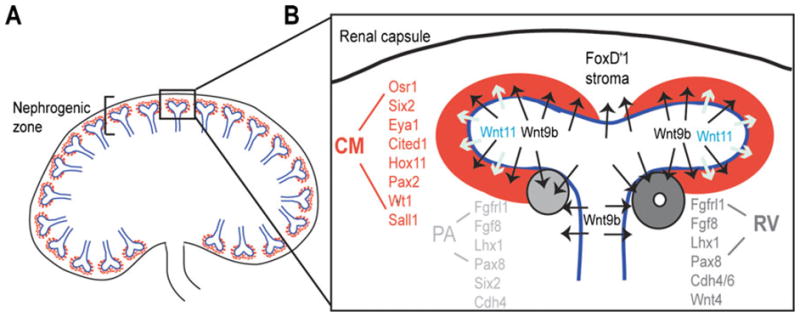
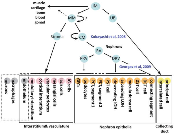
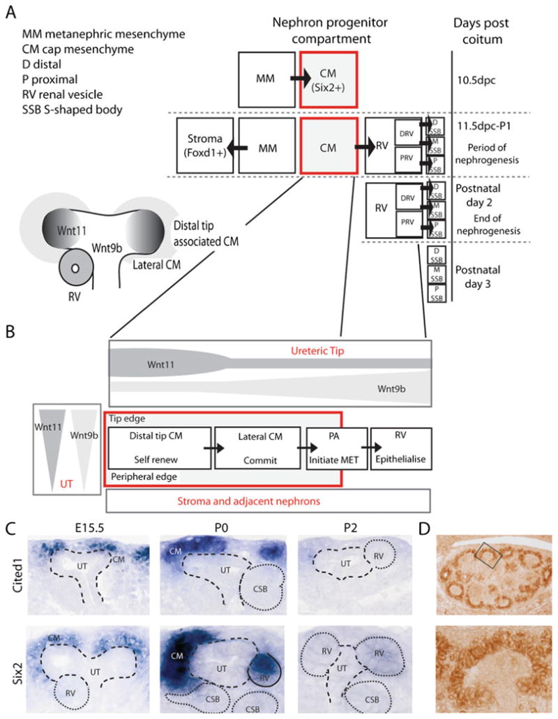
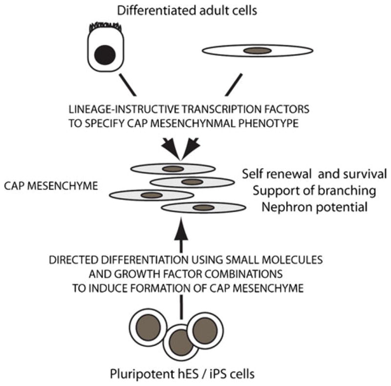
It has long been appreciated that the mammalian kidney arises via reciprocal interactions between an epithelial ureteric epithelium and the surrounding metanephric mesenchyme. More recently, lineage tracing has confirmed that the portion of the metanephric mesenchyme closest to the advancing ureteric tips, the cap mesenchyme, represents the progenitor population for the nephron epithelia. This Six2(+)Cited1(+) population undergoes self-renewal throughout nephrogenesis while retaining the potential to epithelialize. In contrast, the Foxd1(+) portion of the metanephric mesenchyme shows no epithelial potential, developing instead into the interstitial, perivascular, and possibly endothelial elements of the kidney. The cap mesenchyme rests within a nephrogenic niche, surrounded by the stroma and the ureteric tip. While the role of Wnt signaling in nephron induction is known, there remains a lack of clarity over the intrinsic and extrinsic regulation of cap mesenchyme specification, self-renewal, and nephron potential. It is also not known what regulates cessation of nephrogenesis, but there is no nephron generation in response to injury during the postnatal period. In this review, we will examine what is and is not known about this nephron progenitor population and discuss how an increased understanding of the regulation of this population may better explain the observed variation in final nephron number and potentially facilitate the reinitiation or prolongation of nephron formation.
Figures
Similar articles
-
Nephron progenitors in the metanephric mesenchyme.Pediatr Nephrol. 2011 Sep;26(9):1463-7. doi: 10.1007/s00467-011-1806-0. Epub 2011 Feb 19. Pediatr Nephrol. 2011. PMID: 21336811 Review.
-
Osr1 acts downstream of and interacts synergistically with Six2 to maintain nephron progenitor cells during kidney organogenesis.Development. 2014 Apr;141(7):1442-52. doi: 10.1242/dev.103283. Epub 2014 Mar 5. Development. 2014. PMID: 24598167 Free PMC article.
-
Osr1 expression demarcates a multi-potent population of intermediate mesoderm that undergoes progressive restriction to an Osr1-dependent nephron progenitor compartment within the mammalian kidney.Dev Biol. 2008 Dec 1;324(1):88-98. doi: 10.1016/j.ydbio.2008.09.010. Epub 2008 Sep 19. Dev Biol. 2008. PMID: 18835385 Free PMC article.
-
Six2 defines and regulates a multipotent self-renewing nephron progenitor population throughout mammalian kidney development.Cell Stem Cell. 2008 Aug 7;3(2):169-81. doi: 10.1016/j.stem.2008.05.020. Cell Stem Cell. 2008. PMID: 18682239 Free PMC article.
-
Induction and patterning of the metanephric nephron.Semin Cell Dev Biol. 2014 Dec;36:31-8. doi: 10.1016/j.semcdb.2014.08.014. Epub 2014 Sep 3. Semin Cell Dev Biol. 2014. PMID: 25194660 Free PMC article. Review.
Cited by
-
Atlas of Cellular Dynamics during Zebrafish Adult Kidney Regeneration.Stem Cells Int. 2015;2015:547636. doi: 10.1155/2015/547636. Epub 2015 May 18. Stem Cells Int. 2015. PMID: 26089919 Free PMC article.
-
Single-cell multiomics reveals ENL mutation perturbs kidney developmental trajectory by rewiring gene regulatory landscape.Nat Commun. 2024 Jul 15;15(1):5937. doi: 10.1038/s41467-024-50171-w. Nat Commun. 2024. PMID: 39009564 Free PMC article.
-
The number of fetal nephron progenitor cells limits ureteric branching and adult nephron endowment.Cell Rep. 2014 Apr 10;7(1):127-37. doi: 10.1016/j.celrep.2014.02.033. Epub 2014 Mar 20. Cell Rep. 2014. PMID: 24656820 Free PMC article.
-
Cited1 deficiency suppresses intestinal tumorigenesis.PLoS Genet. 2013;9(8):e1003638. doi: 10.1371/journal.pgen.1003638. Epub 2013 Aug 1. PLoS Genet. 2013. PMID: 23935526 Free PMC article.
-
A multicolor podocyte reporter highlights heterogeneous podocyte changes in focal segmental glomerulosclerosis.Kidney Int. 2014 Apr;85(4):972-80. doi: 10.1038/ki.2013.463. Epub 2013 Nov 27. Kidney Int. 2014. PMID: 24284512 Free PMC article.
References
-
- Saxen L, Sariola H. Early organogenesis of the kidney. Pediatr Nephrol. 1987;1:385–392. - PubMed
-
- Georgas KM, Rumballe BA, Valerius MT, Chiu HS, Thiagarajan RD, Lesieur E, Aronow BJ, Brunskill EW, Combes AN, Tang D, Taylor D, Grimmond SM, Potter SS, McMahon AP, Little MH. Analysis of early nephron patterning reveals a role for distal RV proliferation in fusion to the ureteric tip via a cap mesenchyme-derived connecting segment. Dev Biol. 2009;332(2):273–286. - PubMed
-
- Little MH. Regrow or repair – potential regenerative therapies for the kidney. J Am Soc Nephrol. 2006;17(9):2390–2401. - PubMed
Publication types
MeSH terms
Grants and funding
LinkOut - more resources
Full Text Sources
Miscellaneous
