

Natural regulatory T cells control coronary arteriolar endothelial dysfunction in hypertensive mice
- PMID: 21224080
- PMCID: PMC3069876
- DOI: 10.1016/j.ajpath.2010.11.034
Natural regulatory T cells control coronary arteriolar endothelial dysfunction in hypertensive mice
Erratum in
- Am J Pathol. 2011 Mar;178(3):1406. Zakaria, Abd Elmageed [corrected to Abd Elmageed, Zakaria]
Abstract
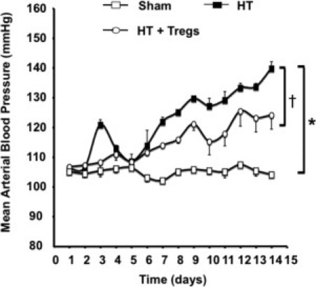
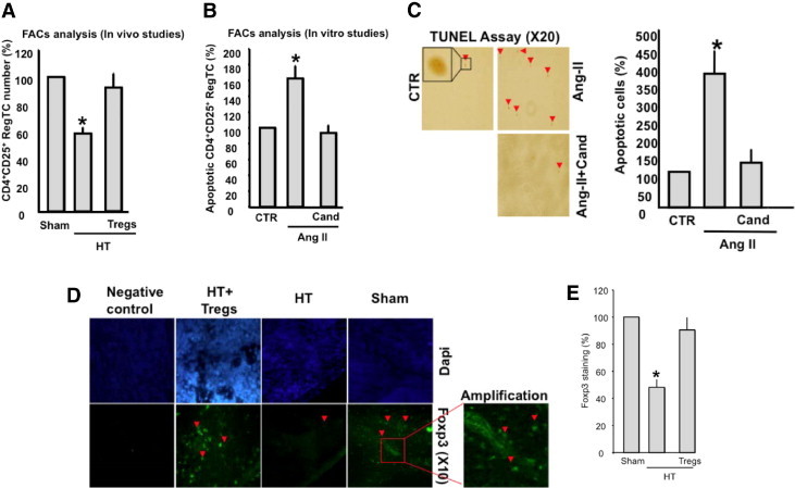
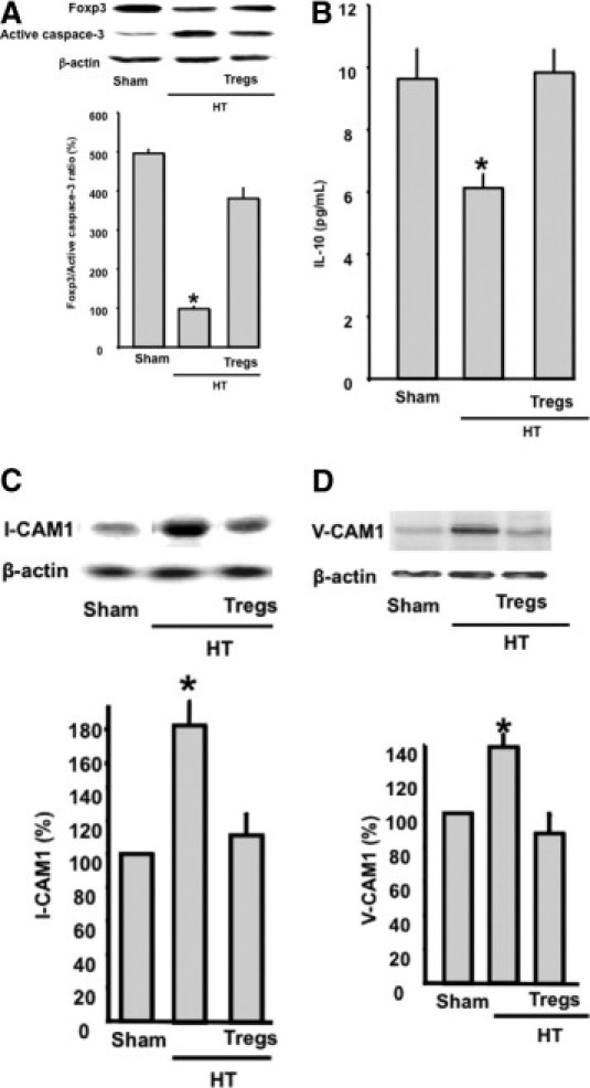
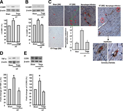
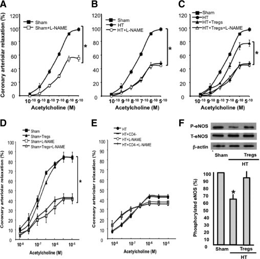
Coronary artery disease in patients with hypertension is increasing worldwide and leads to severe cardiovascular complications. The cellular and molecular mechanisms that underlie this pathologic condition are not well understood. Experimental and clinical research indicates that immune cells and inflammation play a central role in the pathogenesis of cardiovascular diseases. Recently, it has been reported that CD4(+)CD25(+) regulatory T cells (Tregs) regulate heart fibrosis in hypertension. In this study, we determined the role of Tregs in coronary arteriolar endothelial dysfunction in angiotensin II-dependent hypertensive mice. Mice infused with angiotensin II had significantly increased blood pressure, as determined using telemetry, and apoptotic Treg numbers, as measured using flow cytometry. The mice displayed inflammation, assessed by macrophage activation/infiltration into coronary arterioles and the heart, and increased local tumor necrosis factor-α release, which participates in reduced coronary arteriolar endothelial-dependent relaxation in response to acetylcholine using an arteriograph. Hypertensive mice injected with Tregs isolated from control mice had significantly reduced macrophage activation and infiltration, reduced tumor necrosis factor-α release, and improved coronary arteriolar endothelium-dependent relaxation. Our novel data indicate that Tregs are important in the development of coronary arteriolar endothelial dysfunction in hypertension. These results suggest a new direction in the investigation of vascular disease in hypertension and could lead to a therapeutic strategy that involves immune system modulation using Tregs.
Copyright © 2011 American Society for Investigative Pathology. Published by Elsevier Inc. All rights reserved.
Figures
Similar articles
-
T regulatory lymphocytes prevent angiotensin II-induced hypertension and vascular injury.Hypertension. 2011 Mar;57(3):469-76. doi: 10.1161/HYPERTENSIONAHA.110.162941. Epub 2011 Jan 24. Hypertension. 2011. PMID: 21263125
-
Interleukin-10 released by CD4(+)CD25(+) natural regulatory T cells improves microvascular endothelial function through inhibition of NADPH oxidase activity in hypertensive mice.Arterioscler Thromb Vasc Biol. 2011 Nov;31(11):2534-42. doi: 10.1161/ATVBAHA.111.233262. Arterioscler Thromb Vasc Biol. 2011. PMID: 21817097 Free PMC article.
-
Menopause and FOXP3+ Treg cell depletion eliminate female protection against T cell-mediated angiotensin II hypertension.Am J Physiol Heart Circ Physiol. 2019 Aug 1;317(2):H415-H423. doi: 10.1152/ajpheart.00792.2018. Epub 2019 May 17. Am J Physiol Heart Circ Physiol. 2019. PMID: 31099612 Free PMC article.
-
Vascular remodeling and endothelial function in hypertensive patients: effects of antihypertensive therapy.Scand Cardiovasc J Suppl. 1998;47:15-21. doi: 10.1080/140174398428009. Scand Cardiovasc J Suppl. 1998. PMID: 9540129 Review.
-
Role of the endothelium in the genesis of cardiovascular disease.Clin Exp Pharmacol Physiol. 1996 Aug;23(8):S16-22. doi: 10.1111/j.1440-1681.1996.tb03036.x. Clin Exp Pharmacol Physiol. 1996. PMID: 8886508 Review.
Cited by
-
Hypertension: Do Inflammation and Immunity Hold the Key to Solving this Epidemic?Circ Res. 2021 Apr 2;128(7):908-933. doi: 10.1161/CIRCRESAHA.121.318052. Epub 2021 Apr 1. Circ Res. 2021. PMID: 33793336 Free PMC article. Review.
-
The CXCL10/CXCR3 Axis and Cardiac Inflammation: Implications for Immunotherapy to Treat Infectious and Noninfectious Diseases of the Heart.J Immunol Res. 2016;2016:4396368. doi: 10.1155/2016/4396368. Epub 2016 Oct 3. J Immunol Res. 2016. PMID: 27795961 Free PMC article. Review.
-
Developmental endothelial locus-1 protects from hypertension-induced cardiovascular remodeling via immunomodulation.J Clin Invest. 2022 Mar 15;132(6):e126155. doi: 10.1172/JCI126155. J Clin Invest. 2022. PMID: 35133978 Free PMC article.
-
Regulatory T Cells in Chronic Heart Failure.Front Immunol. 2021 Sep 22;12:732794. doi: 10.3389/fimmu.2021.732794. eCollection 2021. Front Immunol. 2021. PMID: 34630414 Free PMC article. Review.
-
ORAI channels in cellular remodeling of cardiorespiratory disease.Cell Calcium. 2019 May;79:1-10. doi: 10.1016/j.ceca.2019.01.005. Epub 2019 Feb 8. Cell Calcium. 2019. PMID: 30772685 Free PMC article. Review.
References
-
- Matrougui K., Maclouf J., Levy B.I., Henrion D. Impaired nitric oxide- and prostaglandin-mediated responses to flow in resistance arteries of hypertensive rats. Hypertension. 1997;30:942–947. - PubMed
-
- Matrougui K., Loufrani L., Levy B.I., Henrion D. High NaCl intake decreases both flow-induced dilation and pressure-induced myogenic tone in resistance arteries from normotensive rats: involvement of cyclooxygenase-2. Pharmacol Toxicol. 2001;89:183–187. - PubMed
-
- Matrougui K., Levy B.I., Schiavi P., Guez D., Henrion D. Indapamide improves flow-induced dilation in hypertensive rats with a high salt intake. J Hypertens. 1998;16:1485–1490. - PubMed
-
- Lominadze D., Joshua I.G., Schuschke D.A. Blood flow shear rates in arterioles of spontaneously hypertensive rats at early and established stages of hypertension. Clin Exp Hypertens. 2001;23:317–328. - PubMed
Publication types
MeSH terms
Grants and funding
LinkOut - more resources
Full Text Sources
Medical
Research Materials
